Comparative Study
. 2005 Mar 30;25(13):3478-87.
doi: 10.1523/JNEUROSCI.3766-04.2005.

Microanatomy of axon/glial signaling during Wallerian degeneration

Affiliations
Comparative Study

Microanatomy of axon/glial signaling during Wallerian degeneration

Amy D Guertin et al. J Neurosci. .

Abstract

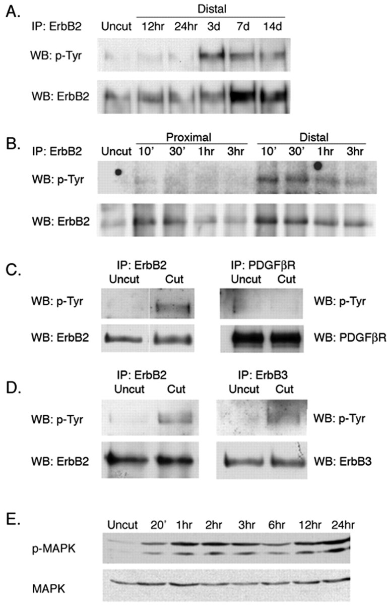
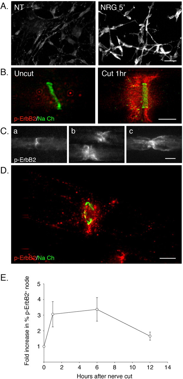
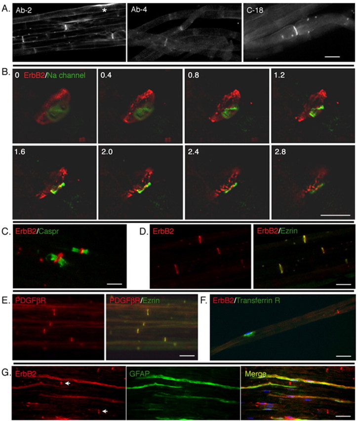
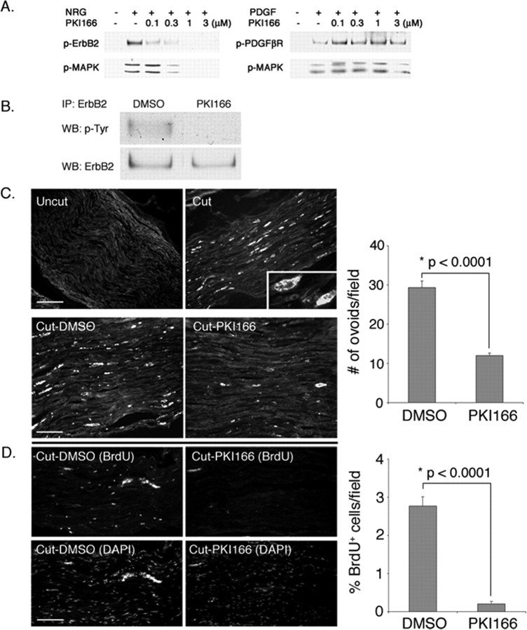
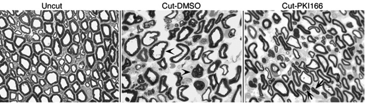
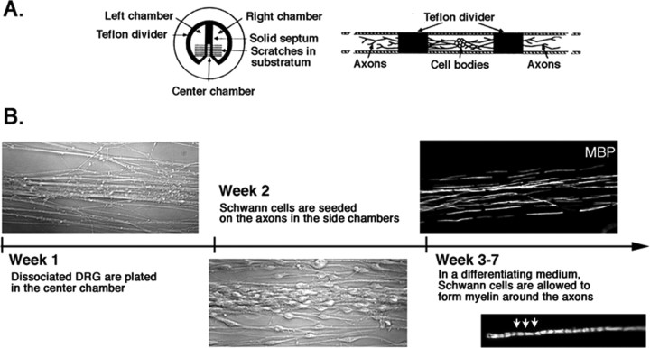
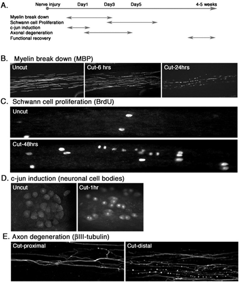
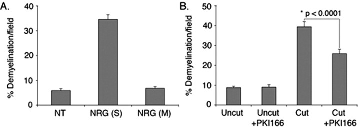
How do myelinated axons signal to the nuclei of cells that enwrap them? The cell bodies of oligodendrocytes and Schwann cells are segregated from axons by multiple layers of bimolecular lipid leaflet and myelin proteins. Conventional signal transduction strategies would seem inadequate to the challenge without special adaptations. Wallerian degeneration provides a model to study axon-to-Schwann cell signaling in the context of nerve injury. We show a hitherto undetected rapid, but transient, activation of the receptor tyrosine kinase erbB2 in myelinating Schwann cells after sciatic nerve axotomy. Deconvolving microscopy using phosphorylation state-specific antibodies shows that erbB2 activation emanates from within the microvilli of Schwann cells, in direct contact with the axons they enwrap. To define the functional role of this transient activation, we used a small molecule antagonist of erbB2 activation (PKI166). The response of myelinating Schwann cells to axotomy is inhibited by PKI166 in vivo. Using neuron/Schwann cell cocultures prepared in compartmentalized cell culture chambers, we show that even transient activation of erbB2 is sufficient to initiate Schwann cell demyelination and that the initiating functions of erbB2 are localized to Schwann cells.


Figures

Figure 1.
Figure 1.
Early and late activation of erbB2 after axotomy. A, Late activation of erbB2. Rat sciatic nerve was transected. At times indicated, the distal stumps were harvested. Nerve lysates were immunoprecipitated with antibodies to erbB2, size fractionated by SDS gel electrophoresis, and immunoblotted with antibodies to p-Tyr or erbB2. As reported previously (Carroll et al., 1997; Kwon et al., 1997), erbB2 protein is overexpressed and also activated at late stages of Wallerian degeneration (3-14 d) at times when Schwann cells have begun to proliferate. IP, Immunoprecipitation; WB, Western blot. B, Early activation of erbB2 (as above, except the nerve stumps were sampled on a timescale of minutes rather than days). As indicated, erbB2 is activated quickly (within 10 min) in the distal stump but not in the proximal stump of axotomized nerves. Activation is maximal between 30 and 60 min after axotomy and is then attenuated. The experiment shown is representative of four replicate experiments. The apparent increase in total erbB seen here at early times in the distal stumps is attributable to experimental variation and is not seen in other experiments. C, Early activation of erbB2 is selective. Nerve lysates from uncut and 1 h postaxotomized distal nerves were immunoprecipitated with erbB2 or PDGFβR antibodies and immunoblotted with p-Tyr antibody. The blots were reused and immunoblotted with erbB2 or PDGFβR antibodies. D, The neuregulin coreceptor erbB3 participates in the early activation response. Nerve lysates were immunoprecipitated with anti-erbB2 or anti-erbB3 and immunoblotted with antibodies to p-Tyr, erbB2, or erbB3, as indicated. As shown, both erbB2 and erbB3 are activated after nerve injury in the distal stump. E, Activation of MAPK in distal stumps after axotomy.
Figure 3.
Figure 3.
Rapid activation of erbB2 at the nodal/paranodal regions in myelinating Schwann cells after nerve injury. A, Immunostaining with phosphospecific antibody detects activation of erbB2. Cultured rat Schwann cells were stimulated with neuregulin (10 ng/ml) for 5 min and then immunostained with a phosphorylation state-specific antibody to erbB2 (p-erbB2). Cells that were left untreated (NT) were used as a control. Positive immunoreactivity is only seen in cells stimulated with neuregulin. Scale bar, 10 μm. B, Rapid activation of erbB2 at the node/paranode regions in myelinating Schwann cells after nerve injury. Teased nerve preparations of an intact nerve (Uncut) and a distal stump (Cut 1 h) were harvested 1 h after axotomy. Nerves were immunostained using p-erbB2 (red) and sodium channel (Na Ch) (green) antibodies to mark the node. Deconvolved microscope images show a punctate pattern of p-erbB2 staining extending outward from the nodal regions of myelinating Schwann cells in cut nerves but not in uncut samples. Scale bar, 5 μm. C, Different patterns of p-erbB2 staining on myelinating Schwann cells after nerve injury. Pictures show p-erbB2 immunostaining on teased nerve fibers prepared 3 h after nerve injury. Activated erbB2 is detected at the nodal (a) and paranodal (b) regions and on the outer membrane of myelinating Schwann cells (c). Scale bar, 10 μm. D, Punctate appearance of p-erbB2 after nerve injury (as in B except the image shows a tangential section through the nodal region of a distal stump Schwann cell at 1 h after axotomy). The node is indicated by immunostaining for sodium channel (green). Puncta of activated erbB2 extend from the nodal region toward the outer plasma membrane of the myelinating Schwann cell. Scale bar, 5 μm. E, Quantitation of erbB2 activation in distal stump using immunostaining. Teased nerve fibers prepared at various hours after axotomy were immunostained for Ezrin and p-erbB2. The total numbers of nodes, marked by Ezrin expression, and p-erbB2-positive nodes and paranodes were counted. Increase in erbB2 activation at each time point was determined by calculating fold increase in percentage of p-erbB2 nodes in axotomized nerve compared with the control (uncut). For each data point, 100-200 nodes were counted. The mean values and SEM were determined from three independent experiments. There is a significant increase in p-erbB2-positive nodes within 1 h after axotomy. This increase is sustained for 6 h and is still discernible (although diminished) at 12 h. In one experiment not shown, we immunostained for p-erbB2 at 24 h after axotomy. At this late time point, no positive p-erbB2 staining was detected.
Figure 2.
Figure 2.
Receptor tyrosine kinases are selectively expressed at the nodal regions of myelinating Schwann cells. A, Low-magnification image of erbB2 expression in rat sciatic nerves. Teased preparations of rat sciatic nerve were immunostained with three different antibody preparations targeted to erbB2 (see Materials and Methods). All three antibodies decorate the nodal regions of the axons. In occasional cells, some perinuclear staining is also seen (*). Scale bar, 10 μm. B, Nodal staining pattern reflects erbB2 in Schwann cells. Deconvolved images of sequential 0.4 μm optical sections (as numbered from top left) of a nodal region of a teased nerve immunostained with antibodies to sodium channel (green) and erbB2 (red). The erbB2 immunofluorescence surrounds the sodium channel, indicating that erbB2 is localized in the nodal region of the myelinating Schwann cell. Scale bar, 5 μm. C, Within Schwann cells, erbB2 is localized to the nodes rather than the paranodes. A teased nerve preparation immunostained with antibodies to erbB2 and Caspr. Expression of erbB2 is flanked by Caspr expression in the paranodes. D, Within the nodes, erbB2 is localized to the microvilli of myelinating Schwann cells. A frozen section of rat sciatic nerve was immunostained with antibodies to erbB2 (red) and Ezrin (green). Expression of erbB2 colocalizes with that of Ezrin, indicating that erbB2 protein is expressed on microvilli of Schwann cells. E, The PDGFβR is also localized to the microvilli [as in D except antibodies were to Ezrin (red) and PDGFβR (green)]. Scale bars: C-E, 15 μm. F, A nutrient transport receptor (transferrin) is targeted to the perinuclear regions of myelinating Schwann cells [as in D and E except that antibodies are directed to the transferrin receptor (green) or erbB2 (red)]. A nucleus of myelinating Schwann cells are indicated by DAPI staining (blue). G, Diffuse expression of erbB2 in nonmyelinating Schwann cells. Frozen sections of rat sciatic nerve were immunostained with antibody against GFAP (green) to visualize nonmyelinating Schwann cells. In GFAP-positive nonmyelinating Schwann cells, erbB2 expression (red) is distributed along the cytoplasm. Nodal expression of erbB2 in myelinating Schwann cells is also shown (arrows). Scale bars: F, G, 30 μm
Figure 4.
Figure 4.
PKI166 blocks Schwann cell response to nerve injury in vivo. A, PKI166 is a selective inhibitor of erbB2 activation. Primary rat Schwann cells were pretreated with various concentrations of PKI166 for 15 min and then stimulated with neuregulin (NRG) (30 ng/ml) or PDGF (30 ng/ml) for 5 min. Cell lysates were prepared and subjected to immunoprecipitation using antibodies to erbB2 or PDGFβR, followed by immunoblotting using antibodies to p-Tyr. Activation of the downstream signaling protein MAPK was determined by immunoblotting using p-MAPK antibody. As indicated, PKI166 inhibits activation of erbB2 and also MAPK in response to neuregulin but has no effect on PDGFβR activation or PDGF-induced signaling events. B, PKI166 inhibits axotomy-induced erbB2 activation in vivo. Thirty minutes before axotomy, rats were injected intraperitoneally with PKI166 or solvent control. One hour after axotomy, distal stump nerves were harvested. Nerve lysates were immunoprecipitated with antibody to erbB2 and immunoblotted with p-Tyr antibody. IP, Immunoprecipitation; WB, Western blot. C, Ovoids of myelin are a convenient metric for demyelination. Frozen sections of uncut or axotomized sciatic nerves were immunostained at 2 d after surgery with antibody to MBP. Top, In uncut nerve, the myelinated axons are evenly stained with the antibody. In a cut nerve, more intense staining on spherical clumps of myelin debris (ovoids) is visible. Inset, Higher magnification of an ovoid stained with anti-MBP antibody. Scale bar, 50 μm. Bottom, Ovoid formation is retarded in PKI166-treated animals. Rats were injected with PKI166 or solvent control as in B. After axotomy, rats were treated with a daily injection of PKI166 (Cut-PKI166) or solvent control (Cut-DMSO) for 2 d. Distal nerves were harvested and immunostained for MBP. Scale bar, 30 μm. D, Schwann cell proliferation is blocked in animals treated with PKI166 after nerve injury. Surgery and drug treatments are the same as in B. Schwann cell proliferation in control and axotomized rats was visualized by injecting rats with BrdU at 2 d after axotomy and then immunostaining with anti-BrdU antibody. Bottom panels show Schwann cell nuclei stained with DAPI. Scale bar, 50 μm. Quantifications of the experiments in C and D are shown on the right. The mean values and SEM were calculated from three independent experiments with each experiment containing data obtained from at least 20-30 random fields. Asterisks indicate statistical significance was determined by ANOVA test.
Figure 5.
Figure 5.
Histology of the sciatic nerves from vehicle- or PKI166-treated rats after nerve injury. Myelin sheaths are visualized by toluidine blue staining on semithin sections of distal sciatic nerves from rats treated with either DMSO or PKI166. Two days after nerve transection, disorganization of myelin sheaths is seen in the DMSO-treated animal (Cut-DMSO), including separation of compact myelin and unwinding of myelin layers (arrowheads). In the PKI166-treated animal (Cut-PKI166), layers of myelin are still compact and smooth with no visible sign of demyelination in most of the Schwann cells. In some cases, compact myelin sheaths are actually retained even in the absence of the axons (arrows).
Figure 6.
Figure 6.
Neuregulin pulse-chase and Schwann cell demyelination in culture. Neuron/Schwann cell cocultures that had myelinated for 4 weeks were treated with neuregulin (30 ng/ml) for various times (30 min, 1, 3, 6, and 48 h), and the culture medium was removed. Cultures were washed with PBS and then incubated continually in fresh medium for the remainder of the experimental period (48 h). Cultures were fixed and immunostained for MBP and scored for demyelination. The mean values and SEM presented for each data point are from at least 80 random fields pooled from a representative experiment. Similar results were observed in triplicate independent experiments.
Figure 7.
Figure 7.
Construction of an in vitro model for Wallerian degeneration using a compartmentalized culture system. A, Compartmental culture chambers. The culture is comprised of three compartments, two side chambers, and a center chamber, separated by silicon-grease-sealed Teflon dividers. DRG sensory neurons are plated in the center compartment, and the axons grow across the divider along the tracks made on the collagen-coated plastic surface. [The illustration is adapted from the study by Campenot and Martin (2001).] B, Formation of myelinated nerves in compartmental cultures. Within 1 week of plating DRG neurons into the center compartment, axons have grown into the side compartments in parallel arrays. At week 2, Schwann cells are plated onto axons in the side chambers. Three to 4 weeks after plating of Schwann cells, myelinated segments on axons are visible by immunostaining using MBP antibodies. Inset, A higher magnification of a myelinating Schwann cell in culture. Schmidth-Lantermann incisors are visible in the internode (arrows).
Figure 8.
Figure 8.
Wallerian degeneration in vitro. A, Time line of Wallerian degeneration in rats. See Results for details. B-E, Wallerian degeneration in culture. Myelinated axons were cut using a razor blade to mimic nerve injury. B, Demyelination in Schwann cells as visualized by immunostaining of the cultures using antibody to MBP. Demyelination in response to nerve injury is detected within 6 h and is well advanced at 24 h. C, Schwann cell proliferation after nerve damage. At 48 h after axotomy, Schwann cells that were formerly in contact with axons began to proliferate as shown by linear arrays of BrdU-positive cells. D, Neuronal responses. Within 1 h after axotomy, the immediate-early gene c-jun protein is visible within the nerve cell bodies in the middle compartment. E, Axonal degeneration in vitro. Within 24 h after axotomy, axons (visualized by immunostaining for β-III tubulin) are distal to the cut degenerate, whereas the proximal axons remain intact.
Figure 9.
Figure 9.
Neuregulin-induced demyelination is a localized effect on Schwann cells. Neuron/Schwann cell cocultures in the compartmentalized culture system had myelinated for 4 weeks. A, Neuregulin (30 ng/ml) was added to Schwann cells in the side compartments (S) or to neuronal soma in the middle compartment (M). Forty-eight hours later, cultures were fixed and immunostained for MBP. A significant increase in demyelination was detected when neuregulin was added to the side compartments but not in the middle compartment compared with the cultures left untreated (NT). The mean values and SEM presented for each data point are from at least 80 random fields pooled from a representative experiment. Similar results were observed in duplicate independent experiments. B, Myelinated axons in the side compartments were pretreated with PKI166 (3 μm) for 20 min followed by a cut to the axons using a razor blade. Twenty-four hours later, cultures were processed for immunostaining as in A. Axotomy-induced Schwann cell demyelination was significantly inhibited by PKI166. The mean values and SEM were calculated from three independent experiments with each experiment containing data obtained from at least 30 random fields. *Statistical significance was determined by ANOVA test.

Similar articles

Cited by

References

    1. Baliga RR, Pimental DR, Zhao YY, Simmons WW, Marchionni MA, Sawyer DB, Kelly RA (1999) NRG-1-induced cardiomyocyte hypertrophy. Role of PI-3-kinase, p70S6K, and MEK-MAPK-RSK. Am J Physiol 277: H2026-H2037. - PubMed
    1. Britsch S, Li L, Kirchhoff S, Theuring F, Brinkmann V, Birchmeier C, Riethmacher D (1998) The ErbB2 and ErbB3 receptors and their ligand, neuregulin-1, are essential for development of the sympathetic nervous system. Genes Dev 12: 1825-1836. - PMC - PubMed
    1. Brockes JP, Fields KL, Raff MC (1979) Studies on cultured rat Schwann cells. I. Establishment of purified populations from cultures of peripheral nerve. Brain Res 165: 105-118. - PubMed
    1. Buonanno A, Fischbach GD (2001) Neuregulin and ErbB receptor signaling pathways in the nervous system. Curr Opin Neurobiol 11: 287-296. - PubMed
    1. Burden S, Yarden Y (1997) Neuregulins and their receptors: a versatile signaling module in organogenesis and oncogenesis. Neuron 18: 847-855. - PubMed

Publication types

MeSH terms