Case Reports
. 2005 May;76(5):833-49.
doi: 10.1086/430134. Epub 2005 Mar 30.

Over- and underdosage of SOX3 is associated with infundibular hypoplasia and hypopituitarism

Affiliations
Case Reports

Over- and underdosage of SOX3 is associated with infundibular hypoplasia and hypopituitarism

Kathryn S Woods et al. Am J Hum Genet. 2005 May.

Abstract

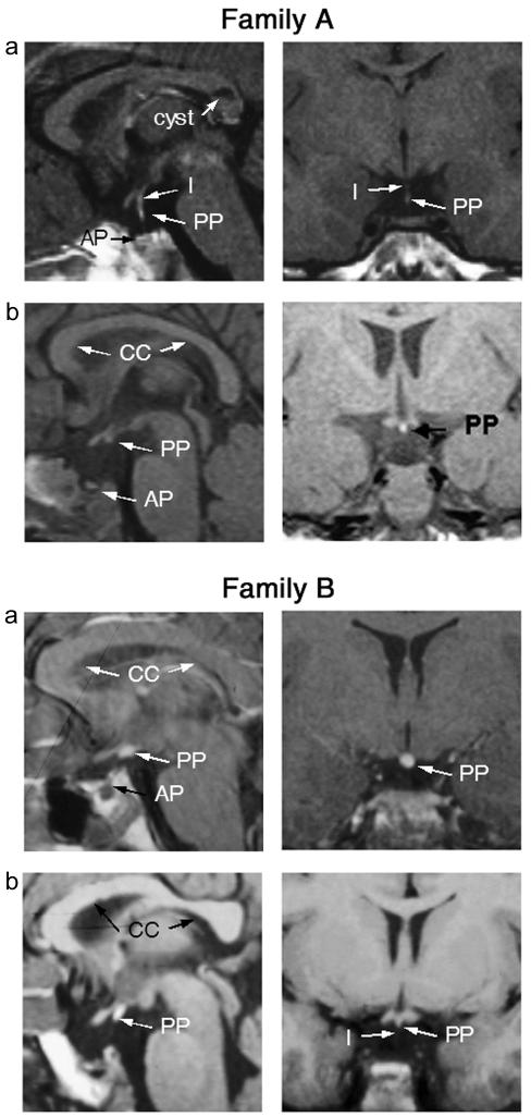
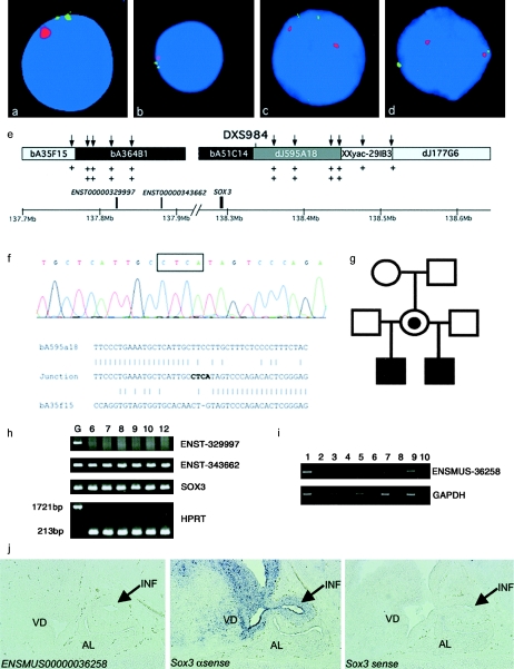
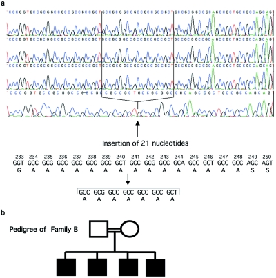
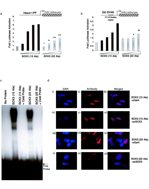
Duplications of Xq26-27 have been implicated in the etiology of X-linked hypopituitarism associated with mental retardation (MR). Additionally, an expansion of a polyalanine tract (by 11 alanines) within the transcription factor SOX3 (Xq27.1) has been reported in patients with growth hormone deficiency and variable learning difficulties. We report a submicroscopic duplication of Xq27.1, the smallest reported to date (685.6 kb), in two siblings with variable hypopituitarism, callosal abnormalities, anterior pituitary hypoplasia (APH), an ectopic posterior pituitary (EPP), and an absent infundibulum. This duplication contains SOX3 and sequences corresponding to two transcripts of unknown function; only Sox3 is expressed in the infundibulum in mice. Next, we identified a novel seven-alanine expansion within a polyalanine tract in SOX3 in a family with panhypopituitarism in three male siblings with an absent infundibulum, severe APH, and EPP. This mutation led to reduced transcriptional activity, with impaired nuclear localization of the mutant protein. We also identified a novel polymorphism (A43T) in SOX3 in another child with hypopituitarism. In contrast to findings in previous studies, there was no evidence of MR or learning difficulties in our patients. We conclude that both over- and underdosage of SOX3 are associated with similar phenotypes, consisting of infundibular hypoplasia and hypopituitarism but not necessarily MR.


Figures

Figure  1
Figure 1
Family A, MRI scans in patients with SOX3 duplication. Coronal and sagittal MRI scans of patient 1 (panel a) and patient 2 (panel b) from family A, showing APH (“AP”), partial hypoplasia of the infundibulum (“I”) in patient 1 (absent in patient 2), and an undescended/ectopic posterior pituitary (“PP”) (partial in patient 1). Note the cyst in the corpus callosum identified in patient 1. Family B, MRI scans in patients with PA expansion in SOX3. Coronal and sagittal MRI scans of patient 2 (panel a) and patient 3 (panel b) from family B, showing APH (“AP”), hypoplasia of the infundibulum (“I”), and an undescended/ectopic posterior pituitary (“PP”).
Figure  2
Figure 2
Duplication of SOX3 in family A. a–d, Representative interphase nuclei from four members of the family (older sibling [a], younger half brother [b], mother [c], and maternal grandmother [d]), with bA51C14 shown in green and the X centromere shown in red. e, Data used to map the extent of the duplication. Clones from the SOX3 genomic region are shown along with position (in Mb) on the X chromosome (NCBI build 34 assembly of the human genome). The clones duplicated by interphase FISH are blackened, those not duplicated are shaded light gray, and the clone containing the breakpoint is shaded dark gray; unknown duplication status is indicated by the unblackened area. The positions of genes within the duplicated region are designated by blackened boxes above the scale bar. The positions of UPQFM-PCR primer pairs are designated by vertical arrows above the contig. Two plus signs underneath a primer pair indicate that dosage quotients for this primer pair were consistent with a duplication; one plus sign shows that this region had a normal copy number. The position of the informative polymorphic marker, DXS984, is also shown. f, Electropherogram showing sequence from the duplication breakpoint in family A. The four nucleotides not present in either the proximal or the distal normal sequence are boxed above the electropherogram. Underneath, a ClustalW alignment of the normal distal (bA595A18), normal proximal (bA35F15), and junction sequence is shown, demonstrating that there is no homology at the proximal and distal ends of the duplication. g, Pedigree of family A. h, RT-PCR for the three genes within the duplication, performed using human brain mRNA at 6, 7, 8, 9, 10, and 12 wk of gestation. Genomic DNA was included as a positive control. Transcripts were present for SOX3 and a novel gene (Ensembl ENST00000343662). The larger 1,721-bp product was not amplified from the brain RNA, showing that there was no genomic DNA contamination. i, RT-PCR analysis for ENSMUSG00000036258 (murine homologue of ENSG00000343662), showing expression in the mouse embryo at 14.5 days postcoitum (dpc). Expression was not detected in the hypothalamic/pituitary region, the pituitary progenitor cell line aT1-1, or the adult pituitary. No amplification was evident in the −RT controls, indicating that the cDNA was not contaminated with genomic DNA. Lane 1, 14.5–dpc embryo +RT; lane 2, 14.5-dpc embryo −RT; lane 3, 14.5-dpc hypothalamus/pituitary +RT; lane 4, 14.5-dpc hypothalamus/pituitary −RT; lane 5, aT1-1 +RT; lane 6, aT1-1 −RT; lane 7, adult pituitary +RT; lane 8, adult pituitary −RT; lane 9, genomic DNA as template; lane 10, water as a blank. j, In situ analysis of the ENSMUSG00000036258 and Sox3 genes in the developing murine hypothalamus, infundibulum, and pituitary. No expression of ENSMUSG00000036258 was detected (left panel). Sox3 is expressed in the infundibulum (arrow) and ventral diencephalon but not in the presumptive anterior pituitary (middle panel). No staining was observed using the Sox3 sense control probe (right panel). INF = infundibulum; VD = ventral diencephalon; AL = anterior lobe.
Figure  3
Figure 3
a, Protein and genomic sequence showing a PA expansion within SOX3. PA expansion (from 15 to 22 alanine residues) in three brothers (top three electropherograms) born to a consanguineous union (family B), compared with the normal wild-type sequence (bottom electropherogram). b, Family tree of a pedigree with a mutation (A(7)240∧241 ins) within SOX3.
Figure  4
Figure 4
a, Impaired activation of Gal4-SOX3(A(7)240∧241 ins) (labeled “SOX3 (22 ala)”) at SOX binding sites. Increasing concentrations of Gal4-SOX3 (labeled “SOX3 (15 ala)”) and Gal4-SOX3(A(7)240∧241 ins) (10 ng, 100 ng, 500 ng, and 1,000 ng) were cotransfected with the HESX1 promoter (Hesx1-PP) luciferase reporter. Gal4-SOX3 led to a dose-dependent activation of the HESX1 promoter (a 6-fold activation at the highest concentration tested), whereas Gal4-SOX3(A(7)240∧241 ins) was associated with impaired activation (a 3-fold activation at the highest concentration tested). b, Impaired activation of Gal4-SOX3(A(7)240∧241 ins) (labeled “SOX3 (22 ala)”) at the SV40 promoter. Increasing concentrations of Gal4-SOX3 (labeled “SOX3 (15 ala)”) and Gal4-SOX3(A(7)240∧241 ins) (10 ng, 100 ng, 500 ng, and 1,000 ng) were cotransfected with the SV40 promoter reporter construct. Gal4-SOX3 led to a dose-dependent activation of the SV40 promoter (a 3.3-fold activation at the highest concentration tested), whereas Gal4-SOX3(A(7)240∧241 ins) was associated with impaired activation (a 2-fold activation at the highest concentration tested). a and b, One asterisk (*) indicates P<.05, two asterisks (**) indicate P<.001, and three asterisks (***) indicate P<.0001. The results represent the means of three independent experiments, each performed in triplicate. c, Gal4-SOX3(A(7)240∧241 ins) (labeled “SOX3 (22 ala)”) binds to SOX binding sites in a manner that is similar to that of wild-type SOX3 (labeled “SOX3 (15 ala)”). Identical amounts of in vitro translated wild-type SOX3 or SOX3(A(7)240∧241 ins) (10 μl) were added to the consensus SOX DNA-binding site (lanes 2 and 4). Duplicate experiments—but with the addition of 15 pmol of cold probe, showing specific loss of DNA binding—are shown in lanes 3 and 5. Lane 1 shows the effect of free probe in the absence of protein. Western blot analysis showed equivalent expression levels of the constructs (data not shown). d, Impaired nuclear localization in Gal4-SOX3(A(7)240∧241) (labeled “SOX3 (22 ala)”). CHO cells were transfected with 50 ng of the respective plasmid construct, as indicated. Nuclei in the cells were counterstained with DAPI. Panels i–vi, wild-type SOX3 (labeled “SOX3 (15 ala)”), using either α-GAL4 (panels i–iii) or α-cSOX3 (panels iv–vi) antibodies; panels vii–xii, Gal4-SOX3(A(7)240∧241) (labeled “SOX3 (22 ala)”), using either α-GAL4 (panels vii–ix) or α-cSOX3 (panels x–xii) antibodies.

References

Electronic-Database Information

    1. Ensembl Genome Browser, http://www.ensembl.org (for ENST00000343662, ENSG00000329997, and ENSMUSG 00000036258)
    1. GenBank, http://www.ncbi.nlm.nih.gov/Genbank/ (for reference sequence [accession number X71135])
    1. Online Mendelian Inheritance in Man (OMIM), http://www.ncbi.nlm.nih.gov/Omim/ (for POU1F1, PROP1, HESX1, LHX3, LHX4, mental retardation, X-linked, with isolated growth hormone deficiency, SOX3, SOX2, FOXL2, HOXA13, HOXD13, PABPN1, RUNX2, ZIC2, ARX, PHOX2, PAX6, SOX9, WNT4, and DAX1)

References

    1. Albrecht AN, Kornak U, Boddrich A, Suring K, Robinson PN, Stiege AC, Lurz R, Stricker S, Wanker EE, Mundlos S (2004) A molecular pathogenesis for transcription factor associated poly-alanine tract expansions. Hum Mol Genet 13:2351–2359 - PubMed
    1. Allen RC, Zoghbi HY, Moseley AB, Rosenblatt HM, Belmont JW (1992) Methylation of HpaII and HhaI sites near the polymorphic CAG repeat in the human androgen-receptor gene correlates with X chromosome inactivation. Am J Hum Genet 51:1229–1239 - PMC - PubMed
    1. Bienvenu T, Poirier K, Friocourt G, Bahi N, Beaumont D, Fauchereau F, Ben Jeema L, Zemni R, Vinet MC, Francis F, Couvert P, Gomot M, Moraine C, Van Bokhoven H, Kalscheuer V, Frints S, Gecz J, Ohzaki K, Chaabouni H, Fryns JP, Desportes V, Beldjord C, Chelly J (2002) ARX, a novel Prd-class-homeobox gene highly expressed in the telencephalon, is mutated in X-linked mental retardation. Hum Mol Genet 11:981–991 - PubMed
    1. Brais B, Bouchard JP, Xie YG, Rochefort DL, Chretien N, Tome FM, Lafreniere RG, Rommens JM, Uyama E, Nohira O, Blumen S, Korczyn AD, Heutink P, Mathieu J, Duranceau A, Codere F, Fardeau M, Rouleau GA, Korcyn AD (1998) Short GCG expansions in the PABP2 gene cause oculopharyngeal muscular dystrophy. Nat Genet 18:164–167 - PubMed
    1. Brickman JM, Adam M, Ptashne M (1999) Interactions between an HMG-1 protein and members of the Rel family. Proc Natl Acad Sci USA 96:10679–10683 - PMC - PubMed

Publication types

MeSH terms