. 2005 Aug 1;389(Pt 3):785-95.
doi: 10.1042/BJ20050337.

Proteomic detection of hydrogen peroxide-sensitive thiol proteins in Jurkat cells

Affiliations

Proteomic detection of hydrogen peroxide-sensitive thiol proteins in Jurkat cells

James W Baty et al. Biochem J. .

Erratum in

  • Biochem J. 2005 Sep 15;390(Pt 3):791-2

Abstract

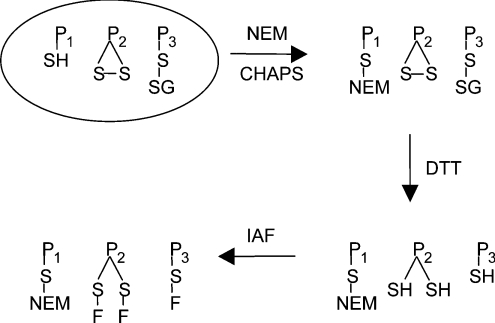
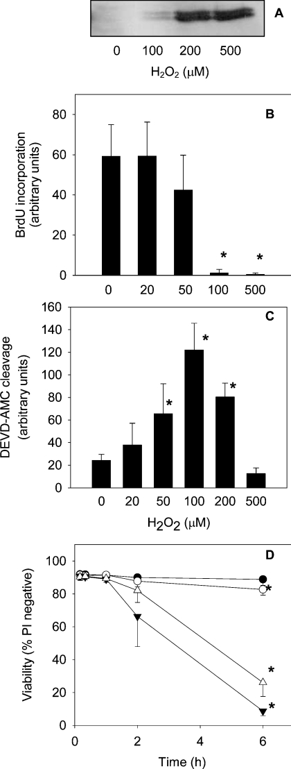
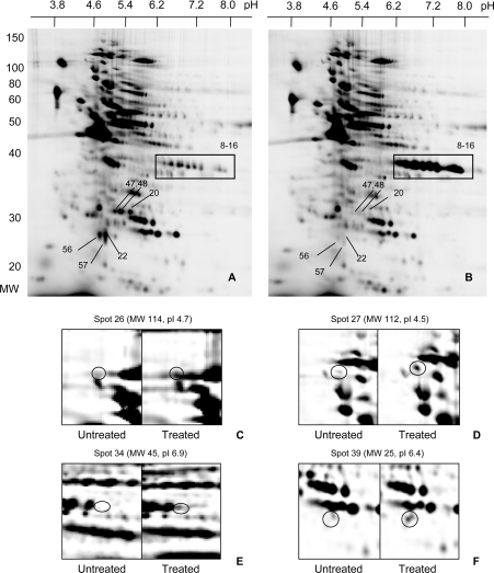
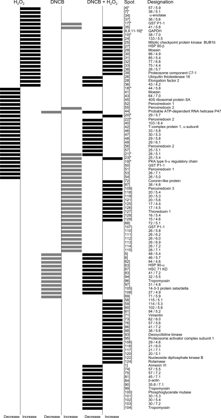
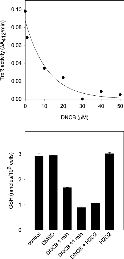
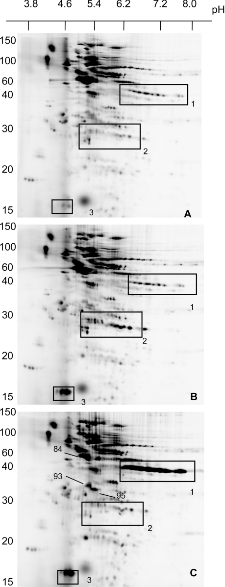
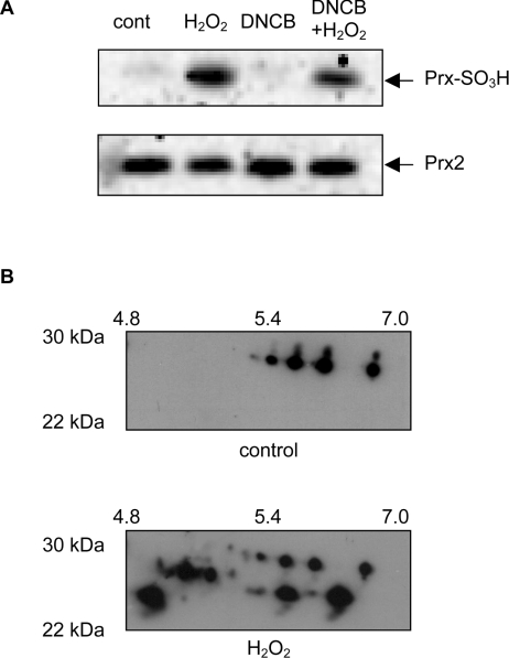
Thiol proteins are important in cellular antioxidant defenses and redox signalling. It is postulated that reactive oxidants cause selective thiol oxidation, but relative sensitivities of different cell proteins and critical targets are not well characterized. We exposed Jurkat cells to H2O2 for 10 min and measured changes in reversibly oxidized proteins by labelling with iodoacetamidofluorescein and two-dimensional electrophoresis. At 200 microM H2O2, which caused activation of the MAP (mitogen-activated protein) kinase ERK (extracellular-signal-regulated kinase), growth arrest and apoptosis, relatively few changes were seen. A total of 28 spots were reversibly oxidized (increased labelling intensity) and 24 decreased. The latter included isoforms of peroxiredoxins 1 and 2, which were irreversibly oxidized. Oxidation of GAPDH (glyceraldehyde-3-phosphate dehydrogenase) was striking, and other affected proteins included glutathione S-transferase P1-1, enolase, a regulatory subunit of protein kinase A, annexin VI, the mitotic checkpoint serine/threonine-protein kinase BUB1beta, HSP90beta (heat-shock protein 90beta) and proteosome components. At 20 microM H2O2, changes were fewer, but GAPDH and peroxiredoxin 2 were still modified. Dinitrochlorobenzene treatment, which inhibited cellular thioredoxin reductase and partially depleted GSH, caused reversible oxidation of several proteins, including thioredoxin 1 and peroxiredoxins 1 and 2. Most changes were distinct from those with H2O2, and changes with H2O2 were scarcely enhanced by dinitrochlorobenzene. Relatively few proteins, including deoxycytidine kinase, nucleoside diphosphate kinase and a proteosome activator subunit, responded only to the combined treatment. Thus most of the effects of H2O2 were not linked to thioredoxin oxidation. Our study has identified peroxiredoxin 2 and GAPDH as two of the most oxidant-sensitive cell proteins and has highlighted how readily peroxiredoxins undergo irreversible oxidation.


Figures

Scheme 1
Scheme 1. Principles for the fluorescence labelling of reversibly oxidized thiol proteins
P1SH represents reduced thiol proteins, P2S-S represents intramolecular disulphides and P3S-SG represents mixed disulphides with glutathione. NEM blocks reduced thiol proteins (P-S-NEM), which are not seen on the gel. DTT reduces reversibly oxidized thiol proteins, which, after treatment with IAF, become fluorescently labelled (P-S-F). Irreversibly oxidized proteins are not labelled. Reversible thiol oxidation gives greater fluorescence labelling of the particular protein spot in oxidant-treated compared with control cells [19].
Figure 1
Figure 1. Jurkat cell responses to H2O2
(A) MAP kinase activity: Jurkat cells (1×10⁶/ml) were treated with H2O2 for 20 min and the dually phosphorylated form of ERK 1/2 was detected in total cell lysates. The Western blot is representative of two independent experiments. (B) Growth arrest: Jurkat cells were treated with H2O2 for 2 h and cell proliferation was measured using the BrdU assay. Results are means±S.D. for at least three experiments. (C) Caspase activation: cells were treated with H2O2 for 6 h and the activity of caspase-3-like enzymes was monitored by cleavage of DEVD-AMC. Data are means and standard deviations from four experiments. (D) Jurkat cell viability as measured by propidium iodide (PI) uptake after treatment with 20 μM H2O2 (●), 200 μM H2O2 (○), 30 μM DNCB (▼) or with a combination of both 30 μM DNCB and 200 μM H2O2 (△). Results are means±S.D. for three experiments. An asterisk (*) indicates a significant difference (P<0.05) when compared with control cells (one-way repeated-measures ANOVA using Bonferroni's method for multiple comparisons).
Figure 2
Figure 2. 2D electrophoresis of oxidized thiol proteins in Jurkat cells
Oxidized thiol proteins from untreated cells (A) and cells treated with 200 μM H2O2 (B) were labelled with IAF and resolved by 2D electrophoresis (10% gel). Boxed regions highlight where major changes were evident. (C)–(F) are enlargements of selected regions showing spots that had undergone major changes. Left-hand panels are from controls and right-hand panels are from treated cells. The numbering of the spots is arbitrary. The identities of all spots that changed, on the basis of molecular mass (MW), pI and, where available, MS analysis, are given in Figure 4 and Table 1.
Figure 3
Figure 3. Cluster diagram summarizing all the thiol-protein changes observed when cells were treated with H2O2, DNCB or with a combination of both DNCB and H2O2
Bars to the left indicate a consistent decrease in IAF labelling intensity on treatment and those to the right indicate an increase. The data shown for H2O2 are with 200 μM H2O2; spots designated * also changed with 20 μM H2O2. There were five spots [numbers 1, 5, 6, and spots at molecular mass 76 kDa/pI 6.1 and at molecular mass 64 kDa/pI 6.2) that increased with 20 μM, but not 200 μM H2O2. The numbering of the spots is arbitrary. Abbreviation: GST, glutathione S-transferase; HSC, heat-shock cognate.
Figure 4
Figure 4. Thioredoxin reductase inhibition (A) and cellular GSH depletion (B) by DNCB
Upper panel: thioredoxin reductase (TrxR) activity after treating cells with DNCB for 10 min. Lower panel: intracellular GSH concentration. Cells were treated with DMSO for 11 min, with 30 μM DNCB for 1 min or 11 min or with 200 μM H2O2 for 10 min. Where cells were exposed to both reagents, the H2O2 was added 1 min after DNCB and incubation was continued for another 10 min. Error bars represent the range of duplicates from one experiment.
Figure 5
Figure 5. 2D electrophoresis of oxidized thiol proteins in control Jurkat cells (A), and cells treated with 30 μM DNCB (B) or with a combination of 200 μM H2O2 and 30 μM DNCB (C)
H2O2 was added 1 min after the addition of DNCB, and cells were incubated for another 10 min. Electrophoresis was performed on 15% gels. Boxes correspond to regions where major changes were evident for GAPDH (box 1), peroxiredoxins (box 2) and thioredoxin (box 3). The values on the extreme left are molecular masses in kDa.
Figure 6
Figure 6. Overoxidation of peroxiredoxins by H2O2
(A) Western blot using a polyclonal antibody that recognizes the sulphinic and sulphonic acid forms of peroxiredoxins 1–3 (Prx-SO3H). Equal loading was confirmed by probing the blots with anti-(peroxiredoxin 2). Jurkat cells were treated with 200 μM H2O2, 30 μM DNCB or a combination of both, and extracted proteins separated by SDS/PAGE. Abbreviation: Prx2, peroxiredoxin 2. (B) Western blot of equivalent regions of 2D gels from control (cont) and 200 μM H2O2-treated cells, showing an increase in the number of bands recognized by the antibody to overoxidized peroxiredoxins.

References

    1. Allen R. G., Tresini M. Oxidative stress and gene regulation. Free Radicals Biol. Med. 2000;28:463–499. - PubMed
    1. Burdon R. H. Superoxide and hydrogen peroxide in relation to mammalian cell proliferation. Free Radicals Biol. Med. 1995;18:775–794. - PubMed
    1. Hampton M. B., Orrenius S. Dual regulation of caspase activity by hydrogen peroxide: implications for apoptosis. FEBS Lett. 1997;414:552–556. - PubMed
    1. Finkel T. Redox-dependent signal transduction. FEBS Lett. 2000;476:52–54. - PubMed
    1. Holmgren A. Antioxidant function of thioredoxin and glutaredoxin systems. Antioxid. Redox Signal. 2000;2:811–820. - PubMed

Publication types

MeSH terms