

IGF-I stimulation of proteoglycan synthesis by chondrocytes requires activation of the PI 3-kinase pathway but not ERK MAPK
- PMID: 15801908
- PMCID: PMC1180722
- DOI: 10.1042/BJ20041636
IGF-I stimulation of proteoglycan synthesis by chondrocytes requires activation of the PI 3-kinase pathway but not ERK MAPK
Abstract
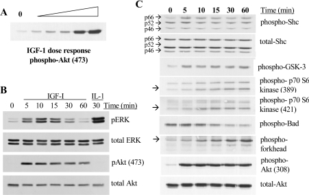
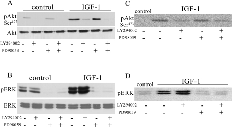
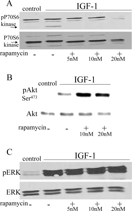
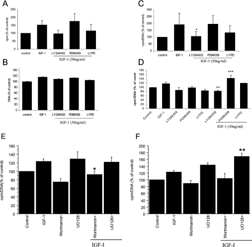
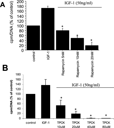
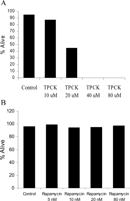
The IGF-I (insulin-like growth factor-I) signalling pathway responsible for regulation of proteoglycan synthesis in chondrocytes has not been defined and is the focus of the present study. Chondrocytes isolated from normal human articular cartilage were stimulated with IGF-I in monolayer culture or in suspension in alginate. IGF-I activated members of both the PI3K (phosphoinositide 3-kinase) pathway and the ERK (extracellular-signal-regulated kinase)/MAPK (mitogen-activated protein kinase) pathway. The PI3K inhibitors LY294002 and wortmannin blocked IGF-I-stimulated Akt phosphorylation without blocking ERK phosphorylation and this was associated with complete inhibition of proteoglycan synthesis. A decrease in IGF-I-stimulated proteoglycan synthesis was also observed upon inhibition of mTOR (mammalian target of rapamycin) and p70S6 kinase, both of which are downstream of Akt. The MEK (MAPK/ERK kinase) inhibitors PD98059 and U0126 blocked IGF-I-stimulated ERK phosphorylation but did not block the phosphorylation of Akt and did not decrease proteoglycan synthesis. Instead, in alginate- cultured chondrocytes, the MEK inhibitors increased IGF-I-stimulated proteoglycan synthesis when compared with cells treated with IGF-I alone. This is the first study to demonstrate that IGF-I stimulation of the PI3K signalling pathway is responsible for the ability of IGF-I to increase proteoglycan synthesis. Although IGF-I also activates the ERK/MAPK pathway, ERK activity is not required for proteoglycan synthesis and may serve as a negative regulator.
Figures
Similar articles
-
Oxidative stress inhibits insulin-like growth factor-I induction of chondrocyte proteoglycan synthesis through differential regulation of phosphatidylinositol 3-Kinase-Akt and MEK-ERK MAPK signaling pathways.J Biol Chem. 2009 Nov 13;284(46):31972-81. doi: 10.1074/jbc.M109.056838. Epub 2009 Sep 17. J Biol Chem. 2009. PMID: 19762915 Free PMC article.
-
Extracellular nicotinamide phosphoribosyltransferase (NAMPT/visfatin) inhibits insulin-like growth factor-1 signaling and proteoglycan synthesis in human articular chondrocytes.Arthritis Res Ther. 2012 Jan 30;14(1):R23. doi: 10.1186/ar3705. Arthritis Res Ther. 2012. PMID: 22289259 Free PMC article.
-
IGF-1-stimulated protein synthesis in oligodendrocyte progenitors requires PI3K/mTOR/Akt and MEK/ERK pathways.J Neurochem. 2009 Jun;109(5):1440-51. doi: 10.1111/j.1471-4159.2009.06071.x. Epub 2009 Mar 28. J Neurochem. 2009. PMID: 19453943
-
Biology and pathology of Rho GTPase, PI-3 kinase-Akt, and MAP kinase signaling pathways in chondrocytes.J Cell Biochem. 2010 Jun 1;110(3):573-80. doi: 10.1002/jcb.22604. J Cell Biochem. 2010. PMID: 20512918 Free PMC article. Review.
-
ERK/MAPK signalling in the developing brain: Perturbations and consequences.Neurosci Biobehav Rev. 2021 Dec;131:792-805. doi: 10.1016/j.neubiorev.2021.10.009. Epub 2021 Oct 8. Neurosci Biobehav Rev. 2021. PMID: 34634357 Review.
Cited by
-
Amelioration of Nicotine-Induced Osteoarthritis by Platelet-Derived Biomaterials Through Modulating IGF-1/AKT/IRS-1 Signaling Axis.Cell Transplant. 2020 Jan-Dec;29:963689720947348. doi: 10.1177/0963689720947348. Cell Transplant. 2020. PMID: 32757664 Free PMC article.
-
Activation of the extracellular-signal-regulated kinase (ERK)/c-Jun N-terminal kinase (JNK) signal pathway and osteogenic factors in subchondral bone of patients with knee osteoarthritis.Ann Transl Med. 2021 Apr;9(8):663. doi: 10.21037/atm-21-1215. Ann Transl Med. 2021. PMID: 33987361 Free PMC article.
-
Berberine ameliorates cartilage degeneration in interleukin-1β-stimulated rat chondrocytes and in a rat model of osteoarthritis via Akt signalling.J Cell Mol Med. 2014 Feb;18(2):283-92. doi: 10.1111/jcmm.12186. Epub 2013 Nov 28. J Cell Mol Med. 2014. PMID: 24286347 Free PMC article.
-
The role of mechano growth factor in chondrocytes and cartilage defects: a concise review.Acta Biochim Biophys Sin (Shanghai). 2023 May 12;55(5):701-712. doi: 10.3724/abbs.2023086. Acta Biochim Biophys Sin (Shanghai). 2023. PMID: 37171185 Free PMC article. Review.
-
Hyperbaric oxygen protects mandibular condylar chondrocytes from interleukin-1β-induced apoptosis via the PI3K/AKT signaling pathway.Am J Transl Res. 2016 Nov 15;8(11):5108-5117. eCollection 2016. Am J Transl Res. 2016. PMID: 27904712 Free PMC article.
References
-
- Jones J. I., Clemmons D. R. Insulin-like growth factors and their binding proteins: biological actions. Endocr. Rev. 1995;16:3–34. - PubMed
-
- Baserga R., Hongo A., Rubini M., Prisco M., Valentinis B. The IGF-I receptor in cell growth, transformation and apoptosis. Biochim. Biophys. Acta. 1997;1332:F105–F126. - PubMed
-
- Powell-Braxton L., Hollingshead P., Warburton C., Dowd M., Pitts-Meek S., Dalton D., Gillett N., Stewart T. A. IGF-I is required for normal embryonic growth in mice. Genes Dev. 1993;7:2609–2617. - PubMed
-
- Liu J. P., Baker J., Perkins A. S., Robertson E. J., Efstratiadis A. Mice carrying null mutations of the genes encoding insulin-like growth factor I (Igf-1) and type 1 IGF receptor (Igf1r) Cell (Cambridge, Mass.) 1993;75:59–72. - PubMed
-
- Butler A. A., Yakar S., Gewolb I. H., Karas M., Okubo Y., LeRoith D. Insulin-like growth factor-I receptor signal transduction: at the interface between physiology and cell biology. Comp. Biochem. Physiol. B Biochem. Mol. Biol. 1998;121:19–26. - PubMed
Publication types
MeSH terms
Substances
Grants and funding
LinkOut - more resources
Full Text Sources
Other Literature Sources
Research Materials
Miscellaneous