

Cellular basis of antiproliferative and antitumor activity of the novel camptothecin derivative, gimatecan, in bladder carcinoma models
- PMID: 15802020
- PMCID: PMC1501124
- DOI: 10.1593/neo.04397
Cellular basis of antiproliferative and antitumor activity of the novel camptothecin derivative, gimatecan, in bladder carcinoma models
Abstract
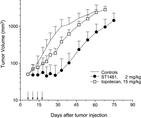
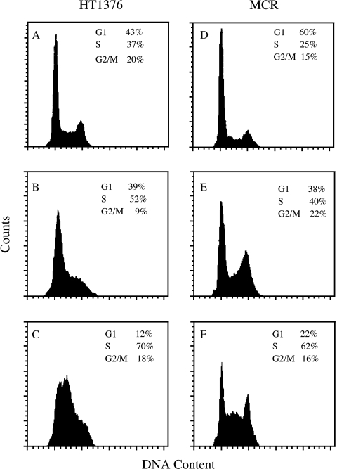
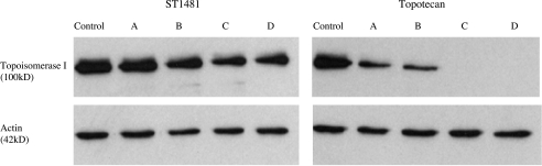
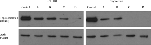
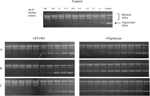
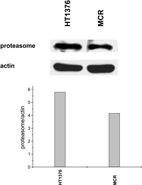
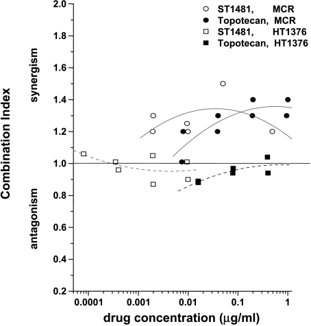
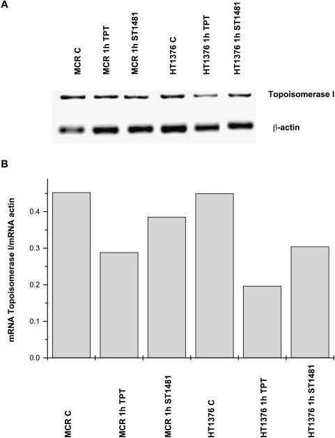
To investigate the cellular/molecular basis of the activity of a novel lipophilic camptothecin, gimatecan (ST1481), against slowly proliferating cells, we performed a comparative study of topotecan and gimatecan in human bladder cancer models (HT1376 and MCR). Gimatecan was significantly more effective than topotecan in inhibiting the growth of HT1376 tumor, thus reflecting antiproliferative potency. In both HT1376 and MCR cells, gimatecan caused a persistent S-phase arrest, indicating an efficient DNA damage checkpoint. This response was consistent with a cytostatic effect, because no evidence of apoptosis was detected. In contrast to gimatecan, topotecan at equitoxic concentrations caused an early and persistent downregulation of topoisomerase I. Modulation of protein level could not be solely ascribed to the proteasome-mediated degradation of the enzyme because the proteasome inhibitor PS341 sensitized MCR but not HT1376 cells to camptothecins, suggesting alternative mechanisms of drug-induced topoisomerase I downregulation. Indeed, the two camptothecins caused a differential inhibition of topoisomerase I transcription, which is more marked in topotecan-treated cells. The HT1376 model was more sensitive to this immediate decrease of mRNA level. Our data document a marked antitumor activity of gimatecan against a bladder carcinoma model. A limited downregulation of topoisomerase I by gimatecan provides additional insights into the cellular basis of drug potency.
Figures

Similar articles
-
A novel oral camptothecin analog, gimatecan, exhibits superior antitumor efficacy than irinotecan toward esophageal squamous cell carcinoma in vitro and in vivo.Cell Death Dis. 2018 May 31;9(6):661. doi: 10.1038/s41419-018-0700-0. Cell Death Dis. 2018. PMID: 29855512 Free PMC article.
-
Effects of drug efflux proteins and topoisomerase I mutations on the camptothecin analogue gimatecan.Invest New Drugs. 2008 Jun;26(3):205-13. doi: 10.1007/s10637-007-9093-0. Epub 2007 Oct 18. Invest New Drugs. 2008. PMID: 17943230
-
Gimatecan and other camptothecin derivatives poison Leishmania DNA-topoisomerase IB leading to a strong leishmanicidal effect.Biochem Pharmacol. 2013 May 15;85(10):1433-40. doi: 10.1016/j.bcp.2013.02.024. Epub 2013 Mar 1. Biochem Pharmacol. 2013. PMID: 23466420
-
An overview of topoisomerase I-targeting agents.Semin Hematol. 1998 Jul;35(3 Suppl 4):3-12. Semin Hematol. 1998. PMID: 9779876 Review.
-
DNA topoisomerase I inhibitors.Cancer Chemother Biol Response Modif. 1997;17:80-113. Cancer Chemother Biol Response Modif. 1997. PMID: 9551210 Review. No abstract available.
Cited by
-
Several genes involved in the JAK-STAT pathway may act as prognostic markers in pancreatic cancer identified by microarray data analysis.Medicine (Baltimore). 2018 Dec;97(50):e13297. doi: 10.1097/MD.0000000000013297. Medicine (Baltimore). 2018. PMID: 30557977 Free PMC article.
-
Gimatecan exerts potent antitumor activity against gastric cancer in vitro and in vivo via AKT and MAPK signaling pathways.J Transl Med. 2017 Dec 13;15(1):253. doi: 10.1186/s12967-017-1360-z. J Transl Med. 2017. PMID: 29237470 Free PMC article.
-
Natural medicines target tumor vascular microenvironment to inhibit tumor.Genes Dis. 2025 Apr 8;12(6):101623. doi: 10.1016/j.gendis.2025.101623. eCollection 2025 Nov. Genes Dis. 2025. PMID: 40821116 Free PMC article. Review.
-
A review of the past, present, and future directions of neoplasia.Neoplasia. 2005 Dec;7(12):1039-46. doi: 10.1593/neo.05793. Neoplasia. 2005. PMID: 16354585 Free PMC article. Review. No abstract available.
-
[Solasonine-induced Apoptosis in Lung Cancer Cell Line H446 and Its Mechanism].Zhongguo Fei Ai Za Zhi. 2015 Jul;18(7):416-21. doi: 10.3779/j.issn.1009-3419.2015.07.05. Zhongguo Fei Ai Za Zhi. 2015. PMID: 26182866 Free PMC article. Chinese.
References
-
- Zunino F, Pratesi G. Camptothecins in clinical development. Exp Opin Invest Drugs. 2004;13:269–284. - PubMed
-
- Pizzolato JF, Saltz LB. The camptothecins. Lancet. 2003;361:2235–2242. - PubMed
-
- Zunino F, Dallavalle S, Laccabue D, Beretta GL, Merlini L, Pratesi G. Current status and perspectives in the development of camptothecins. Curr Pharm Des. 2003;8:2505–2520. - PubMed
-
- Thomas CJ, Rahier NJ, Hecht SM. Camptothecin: current perspectives. Bioorg Med Chem. 2004;12:1585–1604. - PubMed
-
- Pommier Y, Pourquier P, Fan Y, Strumberg D. Mechanism of action of eukaryotic DNA topoisomerase I and drugs targeted to the enzyme. Biochim Biophys Acta. 1998;1400:83–106. - PubMed
Publication types
MeSH terms
Substances
LinkOut - more resources
Full Text Sources
Other Literature Sources
Medical