. 2005 Apr 12;102(15):5530-4.
doi: 10.1073/pnas.0501263102. Epub 2005 Mar 31.

Multipotent nestin-positive, keratin-negative hair-follicle bulge stem cells can form neurons

Affiliations

Multipotent nestin-positive, keratin-negative hair-follicle bulge stem cells can form neurons

Yasuyuki Amoh et al. Proc Natl Acad Sci U S A. .

Abstract

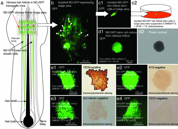
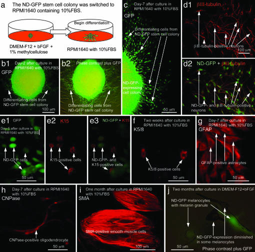
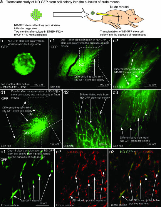
We have recently shown that the expression of nestin, the neural stem cell marker protein, is expressed in bulge-area stem cells of the hair follicle. We used transgenic mice with GFP expression driven by the nestin regulatory element [nestin-driven GFP (ND-GFP)]. The ND-GFP stem cells give rise to the outer-root sheath of the hair follicle as well as an ND-GFP interfollicular vascular network. In this study, we demonstrate that ND-GFP stem cells isolated from the hair-follicle bulge area that are negative for the keratinocyte marker keratin 15 can differentiate into neurons, glia, keratinocytes, smooth muscle cells, and melanocytes in vitro. These pluripotent ND-GFP stem cells are positive for the stem cell marker CD34, as well as keratin 15-negative, suggesting their relatively undifferentiated state. The apparent primitive state of the ND-GFP stem cells is compatible with their pluripotency. Furthermore, we show that cells derived from ND-GFP stem cells can differentiate into neurons after transplantation to the subcutis of nude mice. These results suggest that hair-follicle bulge-area ND-GFP stem cells may provide an accessible, autologous source of undifferentiated multipotent stem cells for therapeutic application.


Figures

Fig. 1.
Fig. 1.
Isolation, culture, and characterization of ND-GFP hair follicle stem cells. (a) Schematic representation of a vibrissa hair follicle in ND-GFP transgenic mice showing the position of the ND-GFP-expressing vibrissa follicular bulge area (red arrows) and ND-GFP-expressing outer-root sheath cells (black arrows). (b) Isolated ND-GFP-expressing vibrissa follicular bulge area contains ND-GFP-expressing hair-follicle stem cells (white arrow heads). (c1 and c2) The ND-GFP-expressing hair-follicle stem cells in the vibrissa follicular bulge area were isolated and suspended in DMEM-F12 containing B-27 and 1% methylcellulose supplemented with bFGF every 2 days. (d1 and d2) After 4 weeks, ND-GFP-expressing hair-follicle stem cells from the vibrissa follicular bulge area formed the ND-GFP-expressing cell colony. (e) ND-GFP-expressing cells within the colony from the vibrissa follicular bulge area were CD34-positive (e1), and the ND-GFP-expressing cells within the colony were K15- (e2), III β-tubulin- (e3), and CD31-negative (e4).
Fig. 2.
Fig. 2.
Differentiation of ND-GFP hair follicle stem cells in vitro. (a) The ND-GFP-expressing cell colony was switched to RPMI medium 1640 containing 10% FBS from DMEM-F12 containing B-27 and 1% methylcellulose supplemented with bFGF every 2 days. (b1 and b2) At 2 days after switching into RPMI medium 1640 containing 10% FBS, differentiating cells migrated out of the ND-GFP-expressing cell colony. (c) At 7 days after switching to RPMI medium 1640, many differentiating cells migrated out of the ND-GFP-expressing cell colony. (d1 and d2) ND-GFP-expressing cells differentiated to III β-tubulin-positive neurons which maintain ND-GFP-expression. (e1e3) At 5 days after switching to RPMI medium 1640, ND-GFP-expressing cells differentiated to K15-positive cells (red fluorescence, arrows) (e2). The K15-positive cells still expressed ND-GFP. (f) ND-GFP-expressing cells differentiated to K5/8-positive cells 2 weeks after switching to RPMI medium 1640. (g) At 7 days after switching to RPMI medium 1640, ND-GFP-expressing cells differentiated to GFAP-positive astrocytes. (h) At 7 days after switching to RPMI medium 1640, ND-GFP-expressing cells differentiated to 2′,3′-cyclic nucleotide 3′-phosphodiesterase (CNPase)-positive oligodendrocytes. (i) At 1 month after culture in RPMI medium 1640 containing 10% FBS, ND-GFP-expressing cells differentiated to SMA-positive smooth muscle cells. (j) At 2 months after culture in DMEM-F12 containing B-27 and 1% methylcellulose supplemented with bFGF every 2 days, ND-GFP-expressing cells differentiated to melanocytes containing melanin. Some melanocytes still expressed ND-GFP.
Fig. 3.
Fig. 3.
Differentiation of ND-GFP hair follicle stem cells after transplantation. ND-GFP-expressing cell colony from the vibrissa follicular bulge area was cultured in DMEM-F12 containing B-27 and 1% methylcellulose supplemented with bFGF every 2 days for 2 months. (a) The ND-GFP-expressing cell colony was transplanted to the subcutis in nude mice. (b) ND-GFP-expressing cell colony before transplantation. (c1 and c2) At 7 days after transplantation into the subcutis in nude mice, ND-GFP-expressing cells migrated from the ND-GFP-expressing cell colony. External image in skin flap. (c2) High magnification of the area in c1 indicated by the white dashed box. (d1d3) At 14 days after transplantation into the subcutis, many ND-GFP-expressing cells migrated from the ND-GFP-expressing cell colony. An external image was made through a skin flap. (d2 and d3) Higher magnification of the areas of d1 indicated by the white dashed boxes. (e) Immunofluorescence staining of III β-tubulin in a frozen section. The ND-GFP-expressing cell colony differentiated to ND-GFP- and III β-tubulin-positive neurons (arrows) (e1e3) in the subcutis of nude mice.

References

    1. Taylor, G., Lehrer, M. S., Jensen, P. J., Sun, T.-T. & Lavker, R. M. (2000) Cell 102, 451–461. - PubMed
    1. Oshima, H., Rochat, A., Kedzia, C., Kobayashi, K. & Barrandon, Y. (2001) Cell 104, 233–245. - PubMed
    1. Tumbar, T., Guasch, G., Greco, V., Blanpain, C., Lowry, W. E., Rendl, M. & Fuchs, E. (2004) Science 303, 359–363. - PMC - PubMed
    1. Morris, R. J., Liu, Y., Marles, L., Yang, Z., Trempus, C., Li, S., Lin, J. S., Sawicki, J. A. & Cotsarelis, G. (2004) Nat. Biotechnol. 22, 411–417. - PubMed
    1. Toma, J. G., Akhavan, M., Fernandes, K. J., Barnabe-Heider, F., Sadikot, A., Kaplan, D. R. & Miller, F. D. (2001) Nat. Cell Biol. 3, 778–784. - PubMed