The crystal structure of a partial mouse Notch-1 ankyrin domain: repeats 4 through 7 preserve an ankyrin fold
- PMID: 15802643
- PMCID: PMC2253258
- DOI: 10.1110/ps.041184105
The crystal structure of a partial mouse Notch-1 ankyrin domain: repeats 4 through 7 preserve an ankyrin fold
Abstract
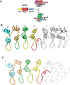
Folding and stability of proteins containing ankyrin repeats (ARs) is of great interest because they mediate numerous protein-protein interactions involved in a wide range of regulatory cellular processes. Notch, an ankyrin domain containing protein, signals by converting a transcriptional repression complex into an activation complex. The Notch ANK domain is essential for Notch function and contains seven ARs. Here, we present the 2.2 A crystal structure of ARs 4-7 from mouse Notch 1 (m1ANK). These C-terminal repeats were resistant to degradation during crystallization, and their secondary and tertiary structures are maintained in the absence of repeats 1-3. The crystallized fragment adopts a typical ankyrin fold including the poorly conserved seventh AR, as seen in the Drosophila Notch ANK domain (dANK). The structural preservation and stability of the C-terminal repeats shed a new light onto the mechanism of hetero-oligomeric assembly during Notch-mediated transcriptional activation.
Figures


References
-
- Artavanis-Tsakonas, S., Rand, M.D., and Lake, R.J. 1999. Notch signaling: Cell fate control and signal integration in development. Science 284 770–776. - PubMed
-
- Barton, G.J. 1993. ALSCRIPT: A tool to format multiple sequence alignments. Protein Eng. 6 37–40. - PubMed
-
- Beatus, P., Lundkvist, J., Oberg, C., and Lendahl, U. 1999. The notch 3 intra-cellular domain represses notch 1-mediated activation through Hairy/Enhancer of split (HES) promoters. Development 126 3925–3935. - PubMed
-
- Beatus, P., Lundkvist, J., Oberg, C., Pedersen, K., and Lendahl, U. 2001. The origin of the ankyrin repeat region in Notch intracellular domains is critical for regulation of HES promoter activity. Mech. Dev. 104 3–20. - PubMed
-
- Bork, P. 1993. Hundreds of ankyrin-like repeats in functionally diverse proteins: Mobile modules that cross phyla horizontally? Proteins 17 363–374. - PubMed
Publication types
MeSH terms
Substances
Grants and funding
LinkOut - more resources
Full Text Sources
Molecular Biology Databases
Research Materials

