Review
. 2005;18(1):41-78.
doi: 10.1089/vim.2005.18.41.

Immune modulation of the hypothalamic-pituitary-adrenal (HPA) axis during viral infection

Affiliations
Review

Immune modulation of the hypothalamic-pituitary-adrenal (HPA) axis during viral infection

Marni N Silverman et al. Viral Immunol. 2005.

Abstract

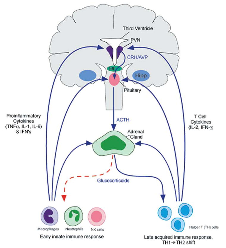
Compelling data has been amassed indicating that soluble factors, or cytokines, emanating from the immune system can have profound effects on the neuroendocrine system, in particular the hypothalamic- pituitary-adrenal (HPA) axis. HPA activation by cytokines (via the release of glucocorticoids), in turn, has been found to play a critical role in restraining and shaping immune responses. Thus, cytokine-HPA interactions represent a fundamental consideration regarding the maintenance of homeostasis and the development of disease during viral infection. Although reviews exist that focus on the bi-directional communication between the immune system and the HPA axis during viral infection (188,235), others have focused on the immunomodulatory effects of glucocorticoids during viral infection (14,225). This review, however, concentrates on the other side of the bi-directional loop of neuroendocrine-immune interactions, namely, the characterization of HPA axis activity during viral infection and the mechanisms employed by cytokines to stimulate glucocorticoid release.


Figures

FIG. 1
FIG. 1
Bidirectional communication between the immune system and the hypothalamic-pituitary-adrenal (HPA) axis (human brain). The immune system, via early innate proinflammatory cytokines (TNFα, IL-1, and IL-6) and interferons, and late acquired T cell cytokines (IL-2 and INF-γ), stimulates glucocorticoid release by acting at all three levels of the HPA axis. In turn, glucocorticoids negatively feedback on the immune system to suppress the further synthesis and release of proinflammatory cytokines (red dotted line). In addition, glucocorticoids play an important role in shaping downtream acquired immune responses, by causing a shift from cellular (Th1/inflammatory) to humoral (Th2/anti-inflammatory) type immune responses. By doing so, glucocorticoids protect an organism from the detrimental consequences of overactive inflammatory immune responses. ACTH, adrenocorticotropic hormone; AVP, arginine vasopressin; CRH, corticotropin-releasing factor; hipp, hippocampus; IFN, interferon: IL, interleukin; ME, median eminence; PVN, paraventricular nucleus of the hypothalamus; TNF, tumor necrosis factor. Reprinted with modifications by permission from Silverman et al. (274).
FIG. 2
FIG. 2
The hypothalamo-pituitary unit. CRH and CRH/AVP neurons originating from the parvocellular division of the PVN of the hypothalamus terminate in the external layer of the median eminence (ME), releasing CRH and or CRH/AVP into the hypophysial-portal circulation, which then act on corticotrophs in the AP to release ACTH into the general circulation. In addition, AVP neurons originating from the magnocellular division of the PVN pass through the internal layer of the ME and terminate on capillaries in the PP to release AVP into the general circulation. ACTH, adrenocorticotropic hormone; AP, anterior pituitary; AVP, arginine vasopressin; CRH, corticotropin-releasing factor; OC, optic chiasm; PP, posterior pituitary; PVN, paraventricular nucleus of the hypothalamus;
FIG. 3
FIG. 3
Cellular and cytokine responses to viral infection and potential immunomodulatory effects of glucocorticoids. Ab, antibody; CTL, cytotoxic T lymphocyte; IFN, interferon; IL, interleukin; NK, natural killer; TNF, tumor necrosis factor; TGF, transforming growth factor. Reprinted by permission from Miller et al. (188).
FIG. 4
FIG. 4
Proposed mechanisms by which peripheral cytokines activate CRH-producing neurons of the PVN in the hypothalamus (rat brain). ACTH, adrenocorticotropic hormone; AP, area postrema; CRH, corticotropin-releasing hormone; HIPP, hippocampus; ME, median eminence; NE, norepenephrine; NO, nitric oxide; NTS, nucleus tractus solitarius; OVLT, organum vascularis of lateral terminalis; PG, prostaglandins; PVN, paraventricular nucleus of the hypothalamus. ★ = site of entry of peripheral cytokine signals. Reprinted by permission from Silverman et al. (274).
FIG. 5
FIG. 5
Local factors regulating glucocorticoid bioavailability and action. (1) corticosterone binding globulin (CBG), (2) 11β-hydroxysteroid dehydrogenase (11β-HSD), (3) multidrug resistance transporter (MDR pump), (4) glucocorticoid receptor (GR = GRα) nuclear translocation, (5) GR interaction with other transcription factors (AP-1 [jun/fos], NFκB), and (6) ratio of GRα:GRβ isoform expression. HSP, heat shock protein; MAP kinase, mitogen-activated protein kinase. Reprinted with modifications by permission from Silverman et al. (274).
FIG. 6
FIG. 6
Kinetics and magnitudes of endogenous glucocorticoid responses vary depending on viral challenge. Serum corticosterone responses to polyinosinic-polycytidylic acid (poly I:C) administration and during viral infections are shown. Mice were injected with 100 μg poly I:C (A) or infected with 2 × 10⁴ PFU of lymphocytic choriomeningitis virus (LCMV) clone E350 (B), 5 × 10⁴ PFU murine cytomegalovirus (MCMV) (C), or 1 × 10⁶ PFU LCMV clone 13 (D). Serum samples were collected under low-stress conditions during the morning (36 h post MCMV-infection) and examined for levels of serum corticosterone. Harvests were at indicated times following treatment or infection. Data are presented as means ± SEM. Reprinted with modifications by permission from Miller et al. (188,189).
FIG. 7
FIG. 7
Interleukin-6 (IL-6) plays a pivotal role in the induction of endogenous glucocorticoids in response to viral challenges. Serum corticosterone levels following murine cytomegalovirus (MCMV), lipopolysaccharide (LPS), polyinosinic-polycytidylic acid (poly I:C), or restraint stress administration in IL-6–deficient and wild-type mice. Corticosterone levels were measured in serum collected from mice under low-stress conditions at 36 h following infection with 5 × 10⁴ PFU MCMV (A); 2 h following injections with PBS, 50 μg LPS, or 100 μg poly I:C (B); or after 30 min of restraint (C). Data are presented as means ± SEM. Results are significant at *p < 0.05. Reprinted by permission from Miller et al. (188,253).
FIG. 8
FIG. 8
Role of glucocorticoids in protection against cytokine (TNF)–mediated lethality. (A) Adrenalectomized (ADX) mice were vehicle injected (squares) or infected with MCMV (1 × 10⁵ PFU/mouse) 5 days following surgery and either not given (circles) or given corticosterone in 0.9% saline as drinking water at either 30 μg/mL (diamonds) or 300 μg/mL (triangles). Control mice received vehicle-treated water. Denoted p value represents significant differences between corticosterone and vehicle-treated MCMV-infected ADX mice. (B) Control (circles) or anti-TNF (triangles) antibodies (Abs) were administered 8–10 h before infection of ADX mice. Denoted p value represents significant differences between TNF and control Ab-treated MCMV-infected ADX mice. Data are presented as means ± SEM. Results are significant at **p < 0.01. Reprinted by permission from Silverman et al. (274,254).
FIG. 9
FIG. 9
Effect of the administration (ip) of a CRH-Ab on low (5 × 10⁴ PFU/mouse) and high (1 × 10⁵ PFU/mouse) dose MCMV-induced plasma ACTH (A), corticosterone (B), and IL-6 (C) responses. Two hundred microliters of either normal sheep serum (NSS) or CRH-antisera was administered 8 h prior to the peak MCMV-induced corticosterone response (28 h post-infection). Trunk blood was collected from C57BL/6 mice at 36 h post-infection (n = 5–11 animals per group). *p < 0.05; **p < 0.01; ***p < 0.001; compared with media/NSS group within dose. #p < 0.05; ##p < 0.01; ###p < 0.001; compared with MCMV/NSS group within dose. One-way ANOVA was used within dose. Because of limited amounts of Ab, a media/CRH-Ab group was not included in this experiment. Reprinted by permission from Silverman et al. (273).
FIG. 10
FIG. 10
Effect of the administration (ip) of an IL-6–Ab on plasma ACTH (A) and corticosterone (B) levels in WT and CRH-KO mice after injection with MCMV (5 × 10⁴ PFU/mouse) or vehicle. The IL-6-Ab (1 mg/mouse) was administered 16 h prior to the peak MCMV-induced corticosterone response (20 h post-infection). Trunk blood was collected from mice at 36 h post-infection (n = 4–8 animals per group). *p < 0.05; ***p < 0.001 (t test for WT/MCMV/IL-6–Ab vs. WT/media/IgG); compared with media/IgG group within genotype. #p < 0.05; ###p < 0.001; compared with MCMV/IgG group within genotype. +p < 0.05; ++p < 0.01 (t test); +++p < 0.001; compared with respective WT group. Two-way ANOVA was used (unless otherwise indicated). Because of limited amounts of Ab, a media/IL-6–Ab group was not included in this experiment. Reprinted by permission from Silverman et al. (273).
FIG. 11
FIG. 11
Plasma ACTH (A), corticosterone (B), and IL-6 (C) levels after injection with MCMV (1 × 10⁵ PFU/mouse) or vehicle in sham-operated or hypophysectomized (hypox) C57BL/6 mice. Trunk blood was collected from mice at 36 h post-infection (n = 3–11 animals per group). ***p < 0.001; compared with media group within surgical-manipulation. ++p < 0.01; +++p < 0.001; compared with respective sham group. Two-way ANOVA indicated a significant interaction of surgery and infection on corticosterone and IL-6 responses for MCMV. Reprinted by permission from Silverman et al. (273).

References

    1. Abbas, A.K., and A.H. Lichtman. 1997. Cellular and Molecular Immunology. Elsevier Science, Philadelphia.
    1. Adcock IM, Brown CR, Barnes PJ. Tumour necrosis factor alpha causes retention of activated glucocorticoid receptor within the cytoplasm of A549 cells. Biochem Biophys Res Commun. 1996;225:545–550. - PubMed
    1. Adcock IM, Caramori G. Cross-talk between pro-inflammatory transcription factors and glucocorticoids. Immunol Cell Biol. 2001;79:376–384. - PubMed
    1. Allan W, Tabi Z, Cleary A, et al. Cellular events in the lymph node and lung of mice with influenza. Consequences of depleting CD4+ T cells J Immunol. 1990;144:3980–3986. - PubMed
    1. Amici C, Belardo G, Rossi A, et al. Activation of I kappa b kinase by herpes simplex virus type 1. A novel target for anti-herpetic therapy. J Biol Chem. 2001;276:28759–28766. - PubMed

MeSH terms