Complement receptors regulate lipopolysaccharide-induced T-cell stimulation
- PMID: 15804286
- PMCID: PMC1782106
- DOI: 10.1111/j.1365-2567.2004.02113.x
Complement receptors regulate lipopolysaccharide-induced T-cell stimulation
Abstract
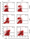
Complement receptors type 1 and 2 (CR1 (CD35)/CR2 (CD21)) are known to enhance the adaptive immune response. In mice, CR1/CR2 are expressed on B cells, follicular dendritic cells, and activated granulocytes. Recently, we showed that a subset of CD44high and CD62Llow T cells also expresses CR1 and CR2. We now report that CR1/CR2 are detectable on both CD4+ and CD8+ subsets of T cells. Lipopolysaccharide (LPS) from Gram-negative bacteria causes polyclonal activation of B cells and stimulation of macrophages and other antigen-presenting cells. We further demonstrate that LPS induced marked up-regulation of CD25 and CD69 on T cells from CR1/CR2 sufficient (Cr+/+), but significantly lower up-regulation on T cells from CR1/CR2 deficient (Cr-/-) mice. These findings point to a novel mechanism by which CR1/CR2 modulates the activation of T cells by LPS.
Figures


Similar articles
-
Cross-linking CD21/CD35 or CD19 increases both B7-1 and B7-2 expression on murine splenic B cells.J Immunol. 1998 Feb 15;160(4):1565-72. J Immunol. 1998. PMID: 9469411
-
Proliferation of resting B cells is modulated by CR2 and CR1.Immunol Lett. 1989 Jun 15;21(4):291-301. doi: 10.1016/0165-2478(89)90022-9. Immunol Lett. 1989. PMID: 2527816
-
Detection of complement receptors 1 and 2 on mouse splenic B cells using flow cytometry.Methods Mol Biol. 2014;1100:305-10. doi: 10.1007/978-1-62703-724-2_24. Methods Mol Biol. 2014. PMID: 24218269
-
Expression and role of CR1 and CR2 on B and T lymphocytes under physiological and autoimmune conditions.Mol Immunol. 2009 Sep;46(14):2767-73. doi: 10.1016/j.molimm.2009.05.181. Epub 2009 Jun 25. Mol Immunol. 2009. PMID: 19559484 Review.
-
Deficiencies of human C3 complement receptors type 1 (CR1, CD35) and type 2 (CR2, CD21).Immunodefic Rev. 1990;2(1):17-41. Immunodefic Rev. 1990. PMID: 2164822 Review.
Cited by
-
App1: an antiphagocytic protein that binds to complement receptors 3 and 2.J Immunol. 2009 Jan 1;182(1):84-91. doi: 10.4049/jimmunol.182.1.84. J Immunol. 2009. PMID: 19109138 Free PMC article.
-
Oocytes could rearrange immunoglobulin production to survive over adverse environmental stimuli.Front Immunol. 2022 Nov 2;13:990077. doi: 10.3389/fimmu.2022.990077. eCollection 2022. Front Immunol. 2022. PMID: 36405746 Free PMC article.
-
Indolent T-cell-rich small B-cell hepatic lymphoma in a Golden Retriever.Clin Case Rep. 2018 Jun 6;6(8):1436-1444. doi: 10.1002/ccr3.1580. eCollection 2018 Aug. Clin Case Rep. 2018. PMID: 30147878 Free PMC article.
-
Endogenous Nur77 Is a Specific Indicator of Antigen Receptor Signaling in Human T and B Cells.J Immunol. 2017 Jan 15;198(2):657-668. doi: 10.4049/jimmunol.1601301. Epub 2016 Dec 9. J Immunol. 2017. PMID: 27940659 Free PMC article.
-
Canine T-zone lymphoma: unique immunophenotypic features, outcome, and population characteristics.J Vet Intern Med. 2014 May-Jun;28(3):878-86. doi: 10.1111/jvim.12343. Epub 2014 Mar 21. J Vet Intern Med. 2014. PMID: 24655022 Free PMC article.
References
-
- Kinoshita T, Takeda J, Hong K, Kozono H, Sakai H, Inoue K. Monoclonal antibodies to mouse complement receptor type 1 (CR1). Their use in a distribution study showing that mouse erythrocytes and platelets are CR1-negative. J Immunol. 1988;140:3066–72. - PubMed
-
- Kaya Z, Afanasyeva M, Wang Y, et al. Contribution of the innate immune system to autoimmune myocarditis: a role for complement. Nat Immunol. 2001;2:739–45. 10.1038/90686. - DOI - PubMed
-
- Molina H, Holers VM, Li B, et al. Markedly impaired humoral immune response in mice deficient in complement receptors 1 and 2. Proc Natl Acad Sci USA. 1996;93:3357–61. 10.1073/pnas.93.8.3357. - DOI - PMC - PubMed
-
- Dempsey PW, Allison ME, Akkaraju S, Goodnow CC, Fearon DT. C3d of complement as a molecular adjuvant: bridging innate and acquired immunity. Science. 1996;271:348–50. - PubMed
-
- Fang Y, Xu C, Fu YX, Holers VM, Molina H. Expression of complement receptors 1 and 2 on follicular dendritic cells is necessary for the generation of a strong antigen-specific IgG response. J Immunol. 1998;160:5273–9. - PubMed
Publication types
MeSH terms
Substances
Grants and funding
LinkOut - more resources
Full Text Sources
Molecular Biology Databases
Research Materials

