Comparative Study
. 2005 Apr 4:6:21.
doi: 10.1186/1471-2202-6-21.

Functional modulation of human delta opioid receptor by neuropeptide FF

Affiliations
Comparative Study

Functional modulation of human delta opioid receptor by neuropeptide FF

Minna-Liisa Ankö et al. BMC Neurosci. .

Abstract

Background: Neuropeptide FF (NPFF) plays a role in physiological pain sensation and opioid analgesia. For example, NPFF potentiates opiate-induced analgesia and the delta opioid receptor antagonist naltrindole inhibits NPFF-induced antinociception. The nature of the interactions between NPFF and opioid receptors seems to be complex and the molecular mechanisms behind the observed physiological effects are not known.

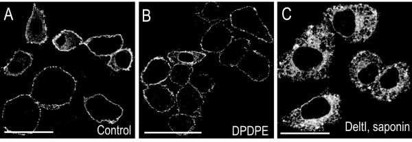
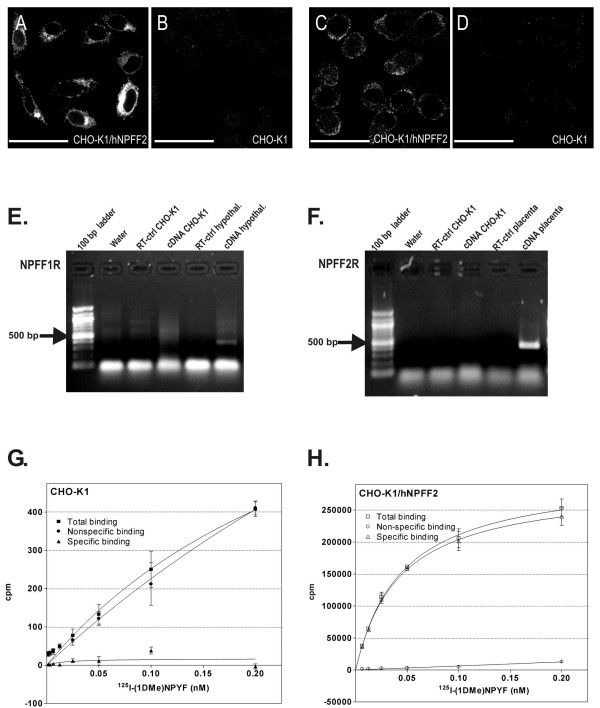
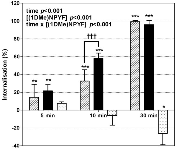
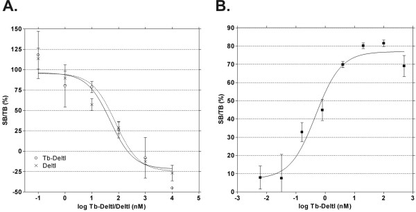
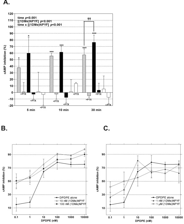
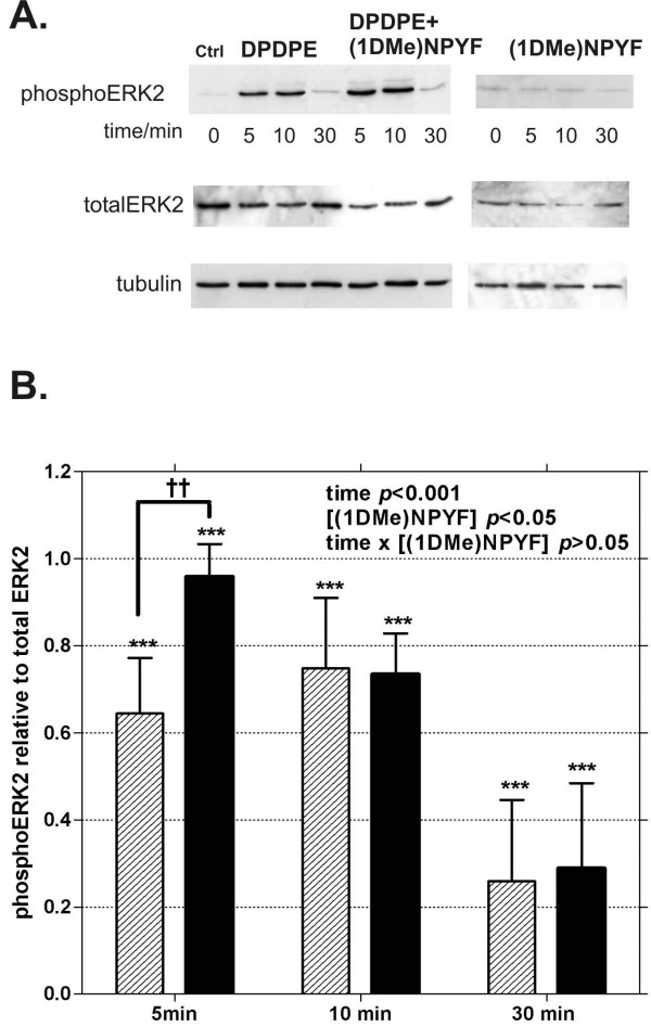
Results: We used a stable Chinese hamster ovary cell line expressing c-MYC-tagged human delta opioid receptor to study the interactions at the molecular level. Our results imply that NPFF can directly modulate the activation of delta opioid receptor in the absence of NPFF receptors. The modulatory effect, though only moderate, was consistently detected with several methods. The agonist-induced receptor trafficking was changed in the presence of (1DMe)NPYF, a stable NPFF-analogue. (1DMe)NPYF enhanced the receptor activation and recovery; opioid antagonists inhibited the effects, indicating that they were delta opioid receptor-mediated. The binding experiments with a novel ligand, Terbium-labeled deltorphin I, showed that (1DMe)NPYF modulated the binding of delta opioid receptor ligands. The levels of phosphorylated mitogen-activated protein kinase and intracellular cAMP were studied to clarify the effects of NPFF on the opioid signaling mechanisms. Application of (1DMe)NPYF together with a delta opioid receptor agonist enhanced the signaling via both pathways studied. Concomitantly to the receptor trafficking, the time-course of the activation of the signaling was altered.

Conclusion: In addition to working via indirect mechanisms on the opioid systems, NPFF may exert a direct modulatory effect on the delta opioid receptor. NPFF may be a multi-functional neuropeptide that regulates several neuronal systems depending on the site of action.


Figures

Figure 1
Figure 1
Characterization of the CHO-K1 cells stably expressing MYC-tagged human DOR. A. The cell surface receptors were visualized with an anti-MYC antibody and a fluorescent secondary antibody. B. The cells were treated with 100 nM DPDPE for 10 min at +37°C after which the remaining cell surface receptors were visualized with an anti-MYC antibody and a fluorescent secondary antibody. C. The cells were stimulated with 100 nM DeltI for 5 min at 37°C, permeabilized with saponin and the receptors were visualized with an anti-MYC antibody and a fluorescent secondary antibody. All the images are 0.5 μm confocal sections at the mid-nuclear level. Representative images are shown. Scale bar 20 μm.
Figure 2
Figure 2
CHO-K1 cells do not express NPFF receptors. The presence or absence of NPFF receptors was analyzed with immunocytochemistry, RT-PCR and radioligand binding assay. A. CHO-K1 cells expressing hNPFF2R showed intense fluorescence when immunolabeled with an anti NPFF2R antibody detecting the C-terminus of the receptor. B. In the same conditions, the C-terminally oriented antibody did not label nontransfected CHO-K1 cells. C. The NPFF2R expression was also studied with an anti-NPFF2R antibody that binds to the N-terminus of the receptor. Again, in CHO-K1 cells expressing hNPFF2R immunoreactivity for the receptor was found. D. The N-terminally oriented NPFF2R antibody did not detect NPFF2R immunoreactivity in the nontransfected CHO-K1 cells. All the images are 0.5 μm confocal sections at the mid-nuclear level. Representative images are shown. Scale bar 20 μm. RT-PCR analysis gave further support for the absence of the NPFF receptors in CHO-K1 cells. E. cDNA from CHO-K1 cells were analyzed with hNPFF1 receptor specific primers and no PCR-product was obtained; human hypothalamus was used as a positive control (expected size of the PCR-product ~350 bp). F. The RT-PCR analysis of CHO-K1 cDNA did not give any PCR-product with hNPFF2R specific primers either; human placenta was used as a positive control (expected size of the PCR-product ~450 bp). G. CHO-K1 cells did not bind the NPFF receptor-specific radioligand at the concentration range where the positive control cell line expressing hNPFF2R showed saturable binding. The solid squares represent the total binding, the solid circles the nonspecific binding and the solid triangles the specific binding to the CHOK1 cell membranes. H. The cell membranes from CHO/hNPFF2R cells were used as a positive control in the experiment. The open squares represent the total binding, the open circles the nonspecific binding and the open triangles the specific binding to the cell membranes.
Figure 3
Figure 3
The quantification of the cell surface fluorescence with FACS after DOR agonist challenge: the effect of (1DMe)NPYF on the agonist- induced internalization of DOR. The cells were first treated with 100 nM DPDPE alone or 100 nM DPDPE + 100 nM (1DMe)NPYF or (1DMe)NPYF at 37°C for indicated times after which the cell surface receptors were detected with an anti-MYC-antibody and a fluorescent secondary antibody. 10000 cells/sample were analyzed with FACS. The combined data of five different experiments performed in duplicates is shown. The data is presented as the internalization percent that is calculated relative to the time-matched control cells (see Experimental section). The statistical significance was analyzed from the non-normalized raw data with two-way ANOVA (Bonferroni's post test, variance as SD, n = 4). *p < 0.05; **p < 0.01 and ***p < 0.001 shows statistical significance relative to the basal level (untreated time-matched cells), †††p < 0.001 significant change in cell surface fluorescence between treatments. The dashed bars represent cells treated with 100 nM DPDPE alone, the solid bars cells treated with 100 nM DPDPE + 100 nM (1DMe)NPYF and the dotted bars represent the cells with 100 nM (1DMe)NPYF alone.
Figure 4
Figure 4
The binding of Tb-DeltI to CHO/MYChDOR -cells. A. Tb-DeltI can displace 3H-diprenorphine with similar affinity as unlabeled DeltI. In the competitive binding experiment 0.1 nM – 10 μM Tb-labeled DeltI or native DeltI was used to displace 6.86 nM3H-diprenorphine from the CHO/MYChDOR cell membranes. Tb-DeltI displaced the radioligand with a similar affinity as the unlabeled native DeltI. The solid line (curve fit) and the crosses represents the competitive binding for DeltI; the dashed line and the open circles for Tb-DeltI. The data is presented as the specific binding (SB) relative to the total binding (TB). The specific binding depicts the radioligand binding that can be displaced by the competing ligand (DeltI or Tb-DeltI). B. Tb-DeltI shows specific, saturable binding to DOR binding sites. The nonspecific binding of Tb-DeltI to CHO/MYChDOR -cells was determined in the presence of 1 μM DeltI. The binding of Tb-DeltI was saturable at a nanomolar range. The data is presented as the specific binding (SB) relative to the total binding (TB). The specific binding depicts the binding of Tb-DeltI that can be displaced by the unlabeled ligand.
Figure 5
Figure 5
(1DMe)NPYF enhances the DOR agonist induced inhibition of forskolin stimulated cAMP accumulation in the CHO-cells expressing MYChDOR. A. The %-cAMP inhibition was calculated relative to the amount of cAMP in the cells treated with only forskolin. The cells were treated at 37°C for the indicated times with 10 μM forskolin together with 100 nM DPDPE (dashed bars), with 100 nM DPDPE + 100 nM (1DMe)NPYF (black solid bars) or with 100 nM (1DMe)NPYF (white solid bars). The bars next to each treatment at each time-point represent the cells pre-treated with pertussis toxin (+PTX). The statistical significance between treatments and time-points is shown in the figure (two-way ANOVA with Bonferroni's post test, variance as SD; significance relative to forskolin only treated cells ***p < 0.001; significance between treatments ††p < 0.01, n = 6). B. The effect of (1DMe)NPYF dose on the DPDPE-induced cAMP-inhibition. The cells were stimulated at 37°C for 30 min with 10 μM forskolin together with 0.1 nM – 10 μM DPDPE and 0 nM – 1 μM (1DMe)NPYF. (1DMe)NPYF caused a significant increase in the DPDPE-induced inhibition of cAMP (two-way Anova, variance as SD; DPDPE p < 0.001; (1DMe)NPYF p < 0.001, [DPDPE] × [(1DMe)NPYF p < 0.01, n = 3). The post-hoc test showed that 10 nM (p < 0.05) and 100 nM (p < 0.05) (1DMe)NPYF had a significant effect on the DPDPE induced cAMP inhibition. The solid circles with thick line represent the cells treated with 0.1 nM – 10 μM DPDPE alone, the open triangles cells treated with DPDPE together with 10 nM (1DMe)NPYF, and the solid squares cells treated with DPDPE together with 100 nM (1DMe)NPYF. C. 1 nM (1DMe)NPYF and 1 μM had only minor effects on the DPDPE induced cAMP inhibition. The solid circles with thick line represent the cells treated with 0.1 nM – 10 μM DPDPE alone, the solid triangles cells treated with DPDPE together with 1 nM (1DMe)NPYF and the open squares DPDPE together with 1 μM (1DMe)NPYF.
Figure 6
Figure 6
DOR agonist induced phosphorylation of the MAP-kinase, ERK2, is increased by (1DMe)NPYF. A. Total cell lysates (10 μg protein per lane) were detected with phosphoERK2 and total ERK2 specific antibodies. The equal loading of samples was controlled with anti-tubulin antibody. The cells were treated with 100 nM DPDPE alone (left panel), with 100 nM DPDPE together with 100 nM (1DMe)NPYF (middle panel) or with 100 nM (1DMe)NPYF (right panel) for 5, 10 or 30 min. (1DMe)NPYF alone could not induce the phosphorylation of ERK2 above the basal level. The data shown are representative figures of five independent experiments. B. The band intensities of phosphoERK2 were quantified and they are presented as relative band intensities with respect to the total ERK2. The dashed bars represent the cells treated with 100 nM DPDPE alone and the solid bars cells treated with 100 nM DPDPE and 100 nM (1DMe)NPYF. The statistical significance between time-points and treatments was analyzed with two-way Anova with Bonferroni's post-test, n = 5. Both of the treatments significantly induced the phosphorylation of the ERK2 above the basal level at all time-points (***p < 0.001). The difference between treatments was significant at the first time-point tested (††p < 0.01).

Similar articles

Cited by

References

    1. Yang H-YT, Fratta W, Majane EA, Costa E. Isolation, sequencing, synthesis, and pharmacological characterization of two brain neuropeptides that modulate the action of morphine. Proc Natl Acad Sci U S A. 1985;82:7757–7761. - PMC - PubMed
    1. Perry SJ, Yi-Kung Huang E, Cronk D, Bagust J, Sharma R, Walker RJ, Wilson S, Burke JF. A human gene encoding morphine modulating peptides related to NPFF and FMRFamide. FEBS Lett. 1997;409:426–430. doi: 10.1016/S0014-5793(97)00557-7. - DOI - PubMed
    1. Vilim FS, Aarnisalo AA, Nieminen M-L, Lintunen M, Karlstedt K, Kontinen VK, Kalso E, States B, Panula P, Ziff E. Gene for pain modulatory neuropeptide NPFF: induction in spinal cord by noxious stimuli. Mol Pharmacol. 1999;55:804–811. - PubMed
    1. Gouarderes C, Jhamandas K, Sutak M, Zajac JM. Role of opioid receptors in the spinal antinociceptive effects of neuropeptide FF analogues. Br J Pharmacol. 1996;117:493–501. - PMC - PubMed
    1. Malin DH, Lake JR, Fowler DE. FMRF-NH2-like mammalian peptide precipitates opiate-withdrawal syndrome in the rat. Peptides. 1990;11:227–280. doi: 10.1016/0196-9781(90)90075-G. - DOI - PubMed

Publication types

Substances

LinkOut - more resources