Comparative Study
. 2005 Jul 1;172(1):85-91.
doi: 10.1164/rccm.200412-1687OC. Epub 2005 Apr 1.

Haemophilus influenzae from patients with chronic obstructive pulmonary disease exacerbation induce more inflammation than colonizers

Affiliations
Comparative Study

Haemophilus influenzae from patients with chronic obstructive pulmonary disease exacerbation induce more inflammation than colonizers

Cecilia L Chin et al. Am J Respir Crit Care Med. .

Abstract

Rationale: Airway infection with Haemophilus influenzae causes airway inflammation, and isolation of new strains of this bacteria is associated with increased risk of exacerbations in patients with chronic obstructive pulmonary disease (COPD).

Objective: To determine whether strains of H. influenzae associated with exacerbations cause more inflammation than strains that colonize the airways of patients with COPD.

Methods: Exacerbation strains of H. influenzae were isolated from patients during exacerbation of clinical symptoms with subsequent development of a homologous serum antibody response and were compared with colonization strains that were not associated with symptom worsening or an antibody response. Bacterial strains were compared using an in vivo mouse model of airway infection and in vitro cell culture model of bacterial adherence and defense gene and signaling pathway activation in primary human airway epithelial cells.

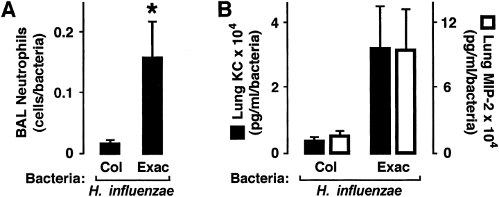
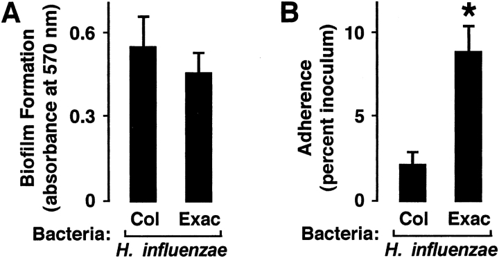
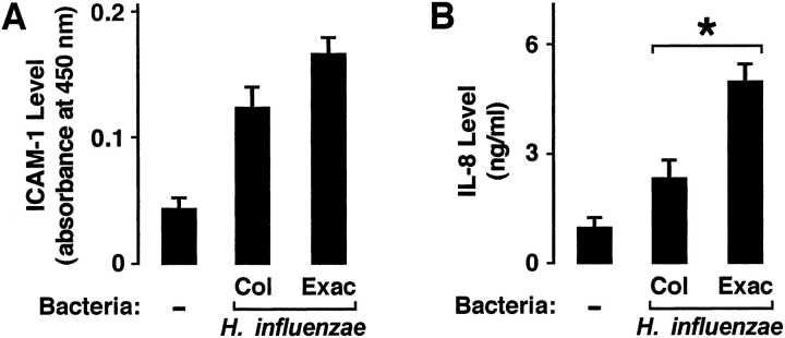
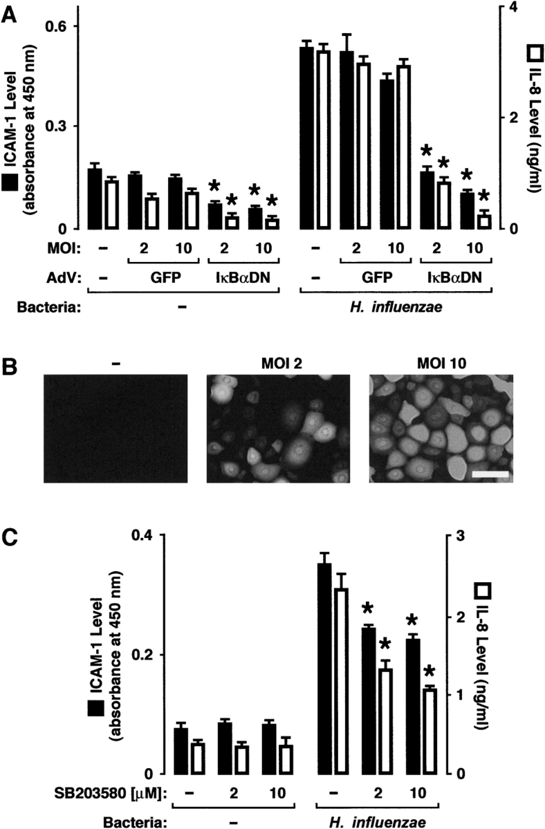
Results: H. influenzae associated with exacerbations caused more airway neutrophil recruitment compared with colonization strains in the mouse model of airway bacterial infection. Furthermore, exacerbation strains adhered to epithelial cells in significantly higher numbers and induced more interleukin-8 release after interaction with airway epithelial cells. This effect was likely mediated by increased activation of the nuclear factor-kappaB and p38 mitogen-activated protein kinase signaling pathways.

Conclusions: The results indicate that H. influenzae strains isolated from patients during COPD exacerbations often induce more airway inflammation and likely have differences in virulence compared with colonizing strains. These findings support the concept that bacteria infecting the airway during COPD exacerbations mediate increased airway inflammation and contribute to decreased airway function.


Figures

<b>Figure 1.</b>
Figure 1.
Haemophilus influenzae from patients with chronic obstructive pulmonary disease (COPD) exacerbation induce more airway inflammation. Bronchoalveolar lavage (BAL) neutrophil numbers (A) and lung keratinocyte chemoattractant (KC) and macrophage-inflammatory protein-2 (MIP-2) levels (B) were determined in C57BL/6J mice after tracheobronchial injection of agar particles suspended with H. influenzae isolates from patients with COPD. At 24 hours after inoculation, the left lung underwent bronchoalveolar lavage with quantification of neutrophils, and the right lung was homogenized for chemokine assay and bacterial quantitation. (A, B) Values are expressed as mean neutrophil number or chemokine concentration per bacterial number ± SEM (n = 3–4 bacterial isolates per condition, with each isolate used to infect two to three individual mice), and a significant difference in levels between animals infected with H. influenzae associated with colonization (Col) versus exacerbation (Exac) is indicated by an asterisk.
<b>Figure 2.</b>
Figure 2.
H. influenzae from patients with COPD with exacerbation adhere more to airway epithelial cells. (A) Bacterial biofilm formation was assessed after adherence of H. influenzae isolates from patients with COPD to polystyrene for 96 hours. Biofilms were stained with crystal violet, nonadherent bacteria were removed by washing, biofilms were dissolved in ethanol, and absorbance at 570 nm was determined. (B) Bacterial adherence to human tracheobronchial epithelial cell monolayers was assessed after incubation of cells for 30 minutes with H. influenzae isolates from patients with COPD. Nonadherent bacteria were removed by washing, cells and bacteria were released by saponin treatment and scraping, and numbers of adherent bacteria were determined by quantitative culture. (A, B) Values are expressed as mean ± SEM (n = 7–10), and a significant difference in bacterial adherence between H. influenzae associated with colonization versus exacerbation is indicated by an asterisk.
<b>Figure 3.</b>
Figure 3.
H. influenzae from patients with COPD with exacerbation induce more interleukin (IL)-8. Intercellular adhesion molecule-1 (ICAM-1) protein expression on the cell surface (A) and IL-8 secretion into the culture media (B) were determined using enzyme-linked immunoassays with hTBE cell monolayers that were incubated for 24 hours without or with equivalent inoculums of H. influenzae isolates from patients with COPD. (A, B) Values are expressed as mean ± SEM (n = 7–10), and a significant difference in IL-8 release induced by H. influenzae associated with colonization versus exacerbation is indicated by an asterisk.
<b>Figure 4.</b>
Figure 4.
H. influenzae induction of epithelial cell ICAM-1 and IL-8 requires nuclear factor κB and p38. (A) ICAM-1 protein expression on the cell surface and IL-8 secretion into the culture media were determined using enzyme-linked immunoassays with hTBE cell monolayers that were left uninfected or were infected with adenoviral vectors expressing control green fluorescence protein (GFP) transgene or dominant-negative IκBα. Cells were then incubated for 24 hours without or with nontypeable H. influenzae strain 12. (B) The proportion of cells expressing transgene protein at each level of infection in (A) was assessed by epifluorescence photomicroscopy with hTBE cell monolayers that were left uninfected or were infected with an adenoviral vector expressing GFP at the indicated MOI. Scale bar = 30 μm. (C) ICAM-1 and IL-8 protein levels were determined using enzyme-linked immunoassays with hTBE cell monolayers that were pretreated with carrier control or a small molecule inhibitor of p38 at the indicated concentrations. Cells were then incubated for 24 hours without or with H. influenzae strain 12. (A,C) Values are expressed as mean ± SEM (n = 3–6), and a significant difference in ICAM-1 expression or IL-8 secretion compared with cells treated with the control is indicated by an asterisk.
<b>Figure 5.</b>
Figure 5.
H. influenzae from patients with COPD with exacerbation activate more NF-κB and p38. (A) NF-κB activation was determined using luciferase assays with hTBE cell monolayers that were initially infected for 24 hours with an adenoviral vector expressing a luciferase gene driven by four tandem NF-κB sites. Cells were then incubated for an additional 24 hours without or with equivalent inoculum of H. influenzae isolates from patients with COPD. (B) Phosphorylation of p38 and extracellular signal–regulated kinases (ERK) mitogen-activated protein (MAP) kinase were assessed using immunoblot analysis of extracts from hTBE cell monolayers that were left uninfected or were infected for 1 hour with equivalent inoculum of H. influenzae isolates from patients with COPD. The positions of phosphorylated and total p38 and ERK are indicated by arrows. (C) Phosphorylation of p38 and ERK that was detected in (B) were quantified using densitometry. (A, C) Values are expressed as mean relative luciferase or phosphorylated/total protein level ± SEM (n = 7–10), and a significant difference in NF-κB or MAP kinase activation induced by H. influenzae associated with colonization versus exacerbation is indicated by an asterisk.

Similar articles

Cited by

References

    1. Burrows B, Earle RH. Course and prognosis of chronic obstructive lung disease: a prospective study of 200 patients. N Engl J Med 1969;280:397–404. - PubMed
    1. Connors AFJ, Dawson NV, Thomas C, Harrell FEJ, Desbiens N, Fulkerson WJ, Kussin P, Bellamy P, Goldman L, Knaus WA. Outcomes following acute exacerbation of severe chronic obstructive lung disease. The SUPPORT investigators (Study to Understand Prognoses and Preferences for Outcomes and Risks of Treatments). Am J Respir Crit Care Med 1996;154:959–967. - PubMed
    1. Seemungal TAR, Donaldson GC, Paul EA, Bestall JC, Jeffries DJ, Wedzicha JA. Effect of exacerbation on quality of life in patients with chronic obstructive pulmonary disease. Am J Respir Crit Care Med 1998;157:1418–1422. - PubMed
    1. Donaldson GC, Seemungal TAR, Bhowmik A, Wedzicha JA. The relationship between exacerbation frequency and lung function decline in chronic obstructive pulmonary disease. Thorax 2002;57:847–852. - PMC - PubMed
    1. Bhowmik A, Seemungal TA, Sapsford RJ, Wedzicha JA. Relation of sputum inflammatory markers to symptoms and lung function changes in COPD exacerbations. Thorax 2000;55:114–120. - PMC - PubMed

Publication types

MeSH terms