

Domain III of Plasmodium falciparum apical membrane antigen 1 binds to the erythrocyte membrane protein Kx
- PMID: 15805191
- PMCID: PMC556269
- DOI: 10.1073/pnas.0501594102
Domain III of Plasmodium falciparum apical membrane antigen 1 binds to the erythrocyte membrane protein Kx
Abstract
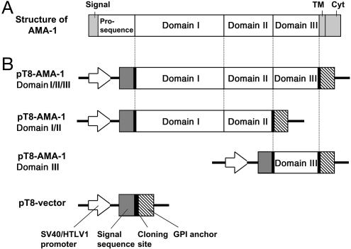
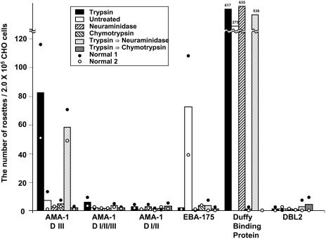
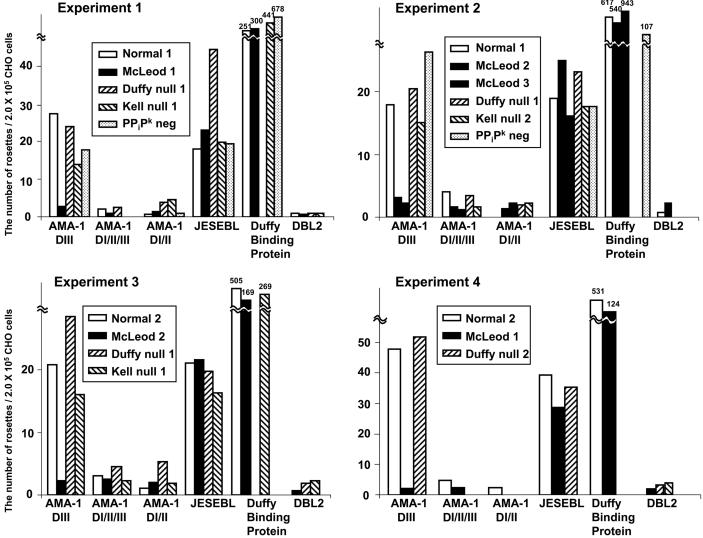
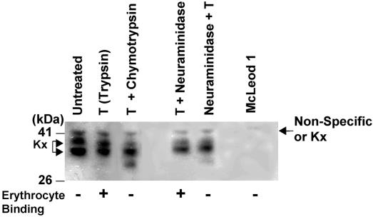
Plasmodium falciparum apical membrane antigen 1 (AMA1) is located in the merozoite micronemes, an organelle that contains receptors for invasion, suggesting that AMA1 may play a role in this process. However, direct evidence that P. falciparum AMA1 binds to human erythrocytes is lacking. In this study, we determined that domain III of AMA1 binds to the erythrocyte membrane protein, Kx, and that the rate of invasion of Kx(null) erythrocytes is reduced, indicating a significant but not unique role of AMA1 and Kx in parasite invasion of erythrocytes. Domains I/II/III, domains I/II and domain III of AMA1 were expressed on the surface of CHO-K1 cells, and their ability to bind erythrocytes was determined. We observed that each of these domains failed to bind untreated human erythrocytes. In contrast, domain III, but not the other domains of AMA1, bound to trypsin-treated human erythrocytes. We tested the binding of AMA1 to trypsin-treated genetically mutant human erythrocytes, missing various erythrocyte membrane proteins. AMA1 failed to bind trypsin-treated Kx(null) (McLeod) erythrocytes, which lack the Kx protein. Furthermore, treatment of human erythrocytes with trypsin, followed by alpha-chymotrypsin, cleaved Kx and destroyed the binding of AMA1 to human erythrocytes. Lastly, the rate of invasion of Kx null erythrocytes by P. falciparum was significantly lower than Kx-expressing erythrocytes. Taken together, our data suggest that AMA1 plays an important, but not exclusive, role in invasion of human erythrocytes through a process that involves exposure or modification of the erythrocyte surface protein, Kx, by a trypsin-like enzyme.
Figures
Similar articles
-
Structures of phage-display peptides that bind to the malarial surface protein, apical membrane antigen 1, and block erythrocyte invasion.Biochemistry. 2003 Aug 26;42(33):9915-23. doi: 10.1021/bi034376b. Biochemistry. 2003. PMID: 12924940
-
Structure and inter-domain interactions of domain II from the blood-stage malarial protein, apical membrane antigen 1.J Mol Biol. 2005 Jul 22;350(4):641-56. doi: 10.1016/j.jmb.2005.05.011. J Mol Biol. 2005. PMID: 15964019
-
Phage-displayed peptides bind to the malarial protein apical membrane antigen-1 and inhibit the merozoite invasion of host erythrocytes.J Biol Chem. 2002 Dec 27;277(52):50303-10. doi: 10.1074/jbc.M207985200. Epub 2002 Oct 14. J Biol Chem. 2002. PMID: 12381731
-
Apical membrane antigen 1 as an anti-malarial drug target.Curr Top Med Chem. 2011;11(16):2039-47. doi: 10.2174/156802611796575885. Curr Top Med Chem. 2011. PMID: 21619512 Review.
-
Vesicle-mediated trafficking of parasite proteins to the host cell cytosol and erythrocyte surface membrane in Plasmodium falciparum infected erythrocytes.Int J Parasitol. 2001 Oct;31(12):1381-91. doi: 10.1016/s0020-7519(01)00256-9. Int J Parasitol. 2001. PMID: 11566305 Review.
Cited by
-
A Presenilin-like protease associated with Plasmodium falciparum micronemes is involved in erythrocyte invasion.Mol Biochem Parasitol. 2008 Mar;158(1):22-31. doi: 10.1016/j.molbiopara.2007.11.007. Epub 2007 Nov 19. Mol Biochem Parasitol. 2008. PMID: 18160114 Free PMC article.
-
A randomized placebo-controlled phase Ia malaria vaccine trial of two virosome-formulated synthetic peptides in healthy adult volunteers.PLoS One. 2007 Oct 10;2(10):e1018. doi: 10.1371/journal.pone.0001018. PLoS One. 2007. PMID: 17925866 Free PMC article. Clinical Trial.
-
Defining species-specific and conserved interactions of apical membrane protein 1 during erythrocyte invasion in malaria to inform multi-species vaccines.Cell Mol Life Sci. 2023 Feb 27;80(3):74. doi: 10.1007/s00018-023-04712-z. Cell Mol Life Sci. 2023. PMID: 36847896 Free PMC article.
-
Extracellular Vesicles Derived from Early and Late Stage Plasmodium falciparum-Infected Red Blood Cells Contain Invasion-Associated Proteins.J Clin Med. 2022 Jul 21;11(14):4250. doi: 10.3390/jcm11144250. J Clin Med. 2022. PMID: 35888014 Free PMC article.
-
Unraveling Haplotype Diversity of the Apical Membrane Antigen-1 Gene in Plasmodium falciparum Populations in Thailand.Korean J Parasitol. 2018 Apr;56(2):153-165. doi: 10.3347/kjp.2018.56.2.153. Epub 2018 Apr 30. Korean J Parasitol. 2018. PMID: 29742870 Free PMC article.
References
-
- Miller, L. H., Baruch, D. I., Marsh, K. & Doumbo, O. K. (2002) Nature 415, 673–679. - PubMed
-
- Bannister, L. H., Hopkins, J. M., Dluzewski, A. R., Margos, G., Williams, I. T., Blackman, M. J., Kocken, C. H., Thomas, A. W. & Mitchell, G. H. (2003) J. Cell Sci. 116, 3825–3834. - PubMed
-
- Triglia, T., Healer, J., Caruana, S. R., Hodder, A. N., Anders, R. F., Crabb, B. S. & Cowman, A. F. (2000) Mol. Microbiol. 38, 706–718. - PubMed
Publication types
MeSH terms
Substances
Grants and funding
LinkOut - more resources
Full Text Sources
Other Literature Sources
