. 2005 May;17(5):1412-23.
doi: 10.1105/tpc.104.030643. Epub 2005 Apr 1.

Molecular and functional dissection of the maize B chromosome centromere

Affiliations

Molecular and functional dissection of the maize B chromosome centromere

Weiwei Jin et al. Plant Cell. 2005 May.

Abstract

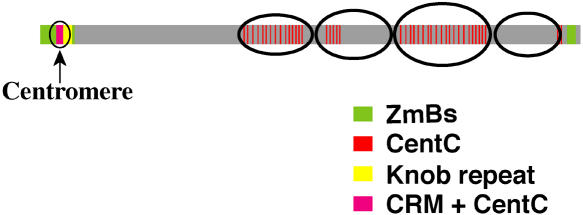
The centromere of the maize (Zea mays) B chromosome contains several megabases of a B-specific repeat (ZmBs), a 156-bp satellite repeat (CentC), and centromere-specific retrotransposons (CRM elements). Here, we demonstrate that only a small fraction of the ZmBs repeats interacts with CENH3, the histone H3 variant specific to centromeres. CentC, which marks the CENH3-associated chromatin in maize A centromeres, is restricted to an approximately 700-kb domain within the larger context of the ZmBs repeats. The breakpoints of five B centromere misdivision derivatives are mapped within this domain. In addition, the fraction of this domain remaining after misdivision correlates well with the quantity of CENH3 on the centromere. Thus, the functional boundaries of the B centromere are mapped to a relatively small CentC- and CRM-rich region that is embedded within multimegabase arrays of the ZmBs repeat. Our results demonstrate that the amount of CENH3 at the B centromere can be varied, but with decreasing amounts, the function of the centromere becomes impaired.


Figures

Figure 1.
Figure 1.
Locations of the CentC, CRM, ZmBs, and Knob Repeats on the Maize Pachytene B Chromosome. Minor sites of these repeats on the B chromosome are not shown. The four distinctive heterochromatin blocks on the long arm are illustrated. The diagram is based on Lamb et al. (2005).
Figure 2.
Figure 2.
Organization of CentC, CRM, and the ZmBs Repeat in the Centromere of Maize B Chromosomes. (A) A fiber-FISH signal derived the ZmBs repeat (green) and CentC (red). Arrows mark the border of the CentC-enriched domain. (B) A diagram based on the fiber-FISH signal pattern in (A). Unambiguous signals from CentC were only observed between the two arrows. (C) A fiber-FISH signal derived the ZmBs repeat (green) and CRM (red). Arrows mark the border of the CentC-enriched domain. (D) A diagram based on the fiber-FISH signal pattern in (C). Additional CRM signals located outside of the CentC-enriched domain are marked by arrowheads. (E) An expanded image of the domain in (A). The five ZmBs repeat arrays are marked. (F) An expanded image of the domain in (C). The five ZmBs repeat arrays are marked. (G) A diagram depicting the distribution patterns of the major blocks of CentC, CRM, and ZmBs. Note: the first ZmBs repeat array contains a distinctive CRM insertion in the middle. Bars = 100 μm in (A) and (C) and 20 μm in (E) and (F).
Figure 3.
Figure 3.
The Pedigree and Structure of the B Centromere Misdivision Derivatives. (A) The pedigree of B centromere misdivision derivatives used in this study. (B) Graphical representation of the centromere size of B chromosome misdivision derivatives based on Kaszas and Birchler (1998). The closed boxes represent the centromeric region.
Figure 4.
Figure 4.
Distribution Patterns of CentC, CRM, and the ZmBs Repeat in the Centromeres of B Centromere Misdivision Derivatives. The ZmBs repeat arrays derived from the CentC-enriched domain are marked. Bars = 30 μm in (E), 25 μm in (A) and (B), 20 μm in (G), 10 μm in (C), (D), (H), and (I), and 5 μm in (F). (A) A fiber-FISH signal of ZmBs (green) and CRM (red) from the centromere of Telo4-11(-). (B) A fiber-FISH signal of ZmBs (green) and CentC (red) from the centromere of Telo4-11(-). (C) A fiber-FISH signal of ZmBs (green) and CentC (red) from the centromere of Ring4-8(-). (D) A fiber-FISH signal of ZmBs (green) and CRM (red) from the centromere of Ring4-8(-). (E) A fiber-FISH signal of ZmBs (green) and CRM (red) from the centromere of Telo4-4(-). (F) A fiber-FISH signal of ZmBs (green) and CRM (red) from the centromere of Ring4-12(-). Note: the ZmBs array is disrupted by a CRM insertion (arrowhead), which is characteristic of the first ZmBs array in the domain. (G) A fiber-FISH signal from ZmBs (green) and a mixed probe of CRM and CentC (red) from the centromere of Telo2-2(-) (fragment 1). (H) A fiber-FISH signal of ZmBs (green) from the centromere of Telo2-2(-) (fragment 2). No CRM and CentC signals were detected within this fragment. (I) A fiber-FISH signal of ZmBs (green) from the centromere of Telo3-3(-). No CRM and CentC signals were detected within this fragment.
Figure 5.
Figure 5.
Diagrams of DNA Organization of Five B Centromere Misdivision Derivatives. All five lines, Telo4-11(-), Telo4-4(-), Ring4-8(-), Ring4-12(-), and Telo2-2(-), contain a DNA fragment derived from the CentC-enriched domain of the original B centromere. The misdivision breakpoints of these five derivatives are all located in the middle of this domain. Vertical arrows point to the borders of the ∼700-kb CentC-enriched domain.
Figure 6.
Figure 6.
Pachytene Localization of the ZmBs and CENH3. (A) Pachytene spreads from Telo2-2(-), Telo4-4(-), and Telo3-3(-) (as an isochromosome in this line) were immunolabeled with CENH3 (in green), hybridized with ZmBs (in red), and counterstained with 4′,6-diamidino-2-phenylindole. (B) Only the ZmBs hybridization signal is shown. The arrow indicates a minor hybridization signal distinct from the main ZmBs hybridization site. The arrowhead indicates a cluster of ZmBs on the distal portion of the reciprocal A-B translocation chromosome in this line. (C) Only the CENH3 signal is shown. Arrows point to the CENH3 signals associated with the misdivided B centromeres. Complete or partial colocalization of ZmBs and CENH3 is always observed for Telo2-2(-) and Telo4-4(-), whereas Telo3-3(-) shows no colocalization in some spreads. (D) A Telo2-2(-) pachytene chromosome labeled with ZmBs (in red) and CentC (in green). Arrow indicates a minor hybridization signal distinct from the main ZmBs hybridization site. Arrowhead indicates the location of CentC hybridization to the B chromosome heterochromatin of the B-A translocation.
Figure 7.
Figure 7.
ChIP Analysis and Cytological Mapping of CENH3 on Stretched Chromatin Fibers and Meiotic and Mitotic Chromosomes. (A) ChIP analysis of CentC and the ZmBs repeat in maize line B73+3B. The ZmBs repeat was coimmunoprecipitated with the anti-CENH3 antibody in all three experiments. The %IP of ZmBs repeat was significantly different from that of the negative control rDNA. (B) A stretched B centromere is stained by the maize anti-CENH3 antibody. The arrows mark the borders of the CENH3 domain. (C) FISH mapping on the same stretched B centromere using the ZmBs repeat. (D) A merged image of the immunostaining and FISH signals. Bar = 10 μm. (E) FISH mapping of CentC (red) and the ZmBs repeat (green) on metaphase I chromosomes of maize B73 line containing a pair of B chromosomes. The Cent repeats (arrows) are located at the most poleward position (ahead of the ZmBs repeats) on the B chromosome bivalent. (F) Immunostaining signals from the anti-CENH3 antibody on the somatic metaphase chromosomes of maize line B73+3B. Arrows point to the signals on the B chromosomes, which are weaker than those on the A centromeres. (G) A merged image of the immunostaining signals with chromosomes, which are stained with 4′,6-diamidino-2-phenylindole and are pseudocolored in red. Bar = 5 μm.
Figure 8.
Figure 8.
Interphase Detection of CENH3 and the ZmBs Repeat in a Maize B73 Line with a Single B Chromosome, Telo4-11(-), Telo4-4(-), Ring4-8(-), and Telo3-3(-). (A) Maize B73 line with a single B chromosome. (B) Telo4-11(-). (C) Telo4-4(-). (D) Ring4-8(-). (E) Telo3-3(-). The signals associated with normal or the misdivided centromeres are indicated by arrows. Note: the ZmBs site in (E) is not associated with any CENH3 staining.

Similar articles

Cited by

References

    1. Alfenito, M.R., and Birchler, J.A. (1993). Molecular characterization of a maize B chromosome centric sequence. Genetics 135, 589–597. - PMC - PubMed
    1. Alonso, A., Mahmood, R., Li, S., Cheung, F., Yoda, K., and Warburton, P.E. (2003). Genomic microarray analysis reveals distinct locations for the CENP-A binding domains in three human chromosome 13q32 neocentromeres. Hum. Mol. Genet. 12, 2711–2721. - PubMed
    1. Amor, D.J., Bentley, K., Ryan, J., Perry, J., Wong, L., Slater, H., and Choo, K.H.A. (2004). Human centromere repositioning “in progress”. Proc. Natl. Acad. Sci. USA 101, 6542–6547. - PMC - PubMed
    1. Ananiev, E.V., Phillips, R.L., and Rines, H.W. (1998). Chromosome-specific molecular organization of maize (Zea mays L.) centromeric regions. Proc. Natl. Acad. Sci. USA 95, 13073–13078. - PMC - PubMed
    1. Carlson, W.R. (1970). Nondisjunction and isochromosome formation in the B chromosome of maize. Chromosoma 30, 356–365.

Publication types

LinkOut - more resources