. 2005 Apr;187(8):2628-37.
doi: 10.1128/JB.187.8.2628-2637.2005.

Flavobacterium johnsoniae GldJ is a lipoprotein that is required for gliding motility

Affiliations

Flavobacterium johnsoniae GldJ is a lipoprotein that is required for gliding motility

Timothy F Braun et al. J Bacteriol. 2005 Apr.

Abstract

Cells of Flavobacterium johnsoniae glide rapidly over surfaces by an unknown mechanism. Eight genes required for gliding motility have been described. Complementation of the nonmotile mutant UW102-48 identified another gene, gldJ, that is required for gliding. gldJ mutants formed nonspreading colonies, and individual cells were completely nonmotile. Like previously described nonmotile mutants, gldJ mutants were deficient in chitin utilization and were resistant to bacteriophages that infect wild-type cells. Cell fractionation and labeling studies with [(3)H]palmitate indicated that GldJ is a lipoprotein. Mutations in gldA, gldB, gldD, gldF, gldG, gldH, or gldI resulted in normal levels of gldJ transcript but decreased levels of GldJ protein. Expression of truncated GldJ protein in wild-type cells resulted in a severe motility defect. GldJ was found in regular bands that suggest the presence of a helical structure within the cell envelope.


Figures

FIG. 1.
FIG. 1.
Map of the gldJ region of F. johnsoniae. Restriction sites are indicated as follows: E, EcoRI; P, PstI; R, EcoRV; X, XbaI. Numbers below the map refer to kilobase pairs of sequence. The presence (+) or absence (−) of complementation of gldJ mutants by fragments cloned into shuttle vectors is indicated beneath the map.
FIG. 2.
FIG. 2.
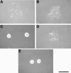
Photomicrographs of F. johnsoniae colonies. Colonies were grown for 30 h at 25°C on PY2 agar medium containing 100 μg of erythromycin/ml. Photomicrographs were taken with a Kodak DC290 digital camera mounted on an Olympus IMT-2 inverted microscope. Bar, 1 mm. (A) Wild-type F. johnsoniae UW101 with shuttle vector pCP11. (B) Wild-type cells with pMM313, which carries gldJ. (C) gldJ mutant UW102-48 with pCP11. (D) UW102-48 complemented with pMM313. (E) Wild-type cells with pMM317, which carries a truncated version of gldJ.
FIG. 3.
FIG. 3.
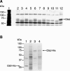
GldJ is a lipoprotein. (A) Cells of F. johnsoniae were labeled with either [3H]glutamate (to label nearly all proteins) or [3H]palmitate (to label lipoproteins). Proteins were separated by SDS-PAGE and detected by autoradiography. Lane 1 contains extracts of wild-type cells labeled with [3H]glutamate, whereas the remaining lanes contain extracts of wild-type, mutant, or complemented cells labeled with [3H]palmitate. Lane 2, wild-type F. johnsoniae. Lane 3, gldB mutant CJ569. Lane 4, CJ569 complemented with pDH233, which carries wild-type gldB. Lane 5, gldD mutant CJ282. Lane 6, CJ282 complemented with pMM213, which carries wild-type gldD. Lane 7, gldH mutant CJ1043. Lane 8, CJ1043 complemented with pMM293, which carries wild-type gldH. Lane 9, gldI mutant UW102-41. Lane 10, UW102-41 complemented with pMM291, which carries wild-type gldI. Lane 11, gldJ mutant UW102-48. Lane 12, UW102-48 complemented with pMM265, which carries wild-type gldJ. (B) Radiolabeling of GldI-His and GldJ-His. Cells of F. johnsoniae were labeled with [3H]palmitate or [3H]glutamate. Proteins were isolated by precipitation with Ni-NTA His-Bind resin, separated by SDS-PAGE, and detected by autoradiography. Lane 1, cells expressing GldI-His from pTB45, labeled with [3H]palmitate. Lane 2, cells expressing GldI-His from pTB45, labeled with [3H]glutamate. Lane 3, cells expressing GldJ-His from pTB44, labeled with [3H]palmitate. Lane 4, cells expressing GldJ-His from pTB44, labeled with [3H]glutamate.
FIG. 4.
FIG. 4.
Immunodetection of GldJ. (A) Whole-cell extracts were examined for GldJ by Western blot analysis. Lane 1, wild-type F. johnsoniae. Lane 2, gldJ mutant UW102-48. Lane 3, UW102-48 with pMM313, which carries gldJ. Equal amounts of protein were loaded in each lane. (B) Cell fractions of wild-type F. johnsoniae were examined for GldJ by Western blot analysis. Lane 1, whole cells. Lane 2, soluble fraction. Lane 3, membrane fraction. Equal amounts of protein were loaded in each lane.
FIG. 5.
FIG. 5.
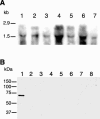
Effect of gld mutations on levels of gldJ mRNA and GldJ protein. (A) Northern blot analysis. RNA was isolated from wild-type and mutant cells and probed with digoxigenin-labeled gldJ RNA. Lane 1, wild-type F. johnsoniae UW101. Lane 2, gldA mutant CJ101-288. Lane 3, gldB mutant CJ569. Lane 4, gldD mutant CJ282. Lane 5, gldFG mutant CJ787. Lane 6, gldH mutant CJ1043. Lane 7, gldI mutant UW102-41. Equal amounts of RNA were loaded in each lane. (B) Western blot analysis of whole-cell extracts with the use of antiserum to GldJ. Lane 1, wild-type F. johnsoniae UW101. Lane 2, gldJ mutant UW102-80, Lane 3, gldA mutant CJ101-288. Lane 4, gldB mutant CJ569. Lane 5, gldD mutant CJ282. Lane 6, gldFG mutant CJ787. Lane 7, gldH mutant CJ1043. Lane 8, gldI mutant UW102-41. Equal amounts of protein were loaded in each lane.
FIG. 6.
FIG. 6.
Localization of GldJ by immunofluorescence microscopy and transmission electron microscopy. Cells of F. johnsoniae were fixed with 1% formaldehyde for 15 min and permeabilized with 5 mM EDTA and 2% Triton X-100, and GldJ was detected using affinity-purified antiserum. Cells were observed by confocal fluorescence microscopy (A) or by transmission electron microscopy (B to D). (A) Immunofluorescent image of wild-type cells incubated with antiserum to GldJ. (B) Electron microscopic image of wild-type cells incubated with antiserum to GldJ. (C) Wild-type cells undergoing cell division incubated with antiserum to GldJ. (D) Cells of the gldJ mutant UW102-48 incubated with antiserum to GldJ. Arrows in panel B indicate cells exhibiting labeling of helical structures. Large arrowheads in panels B and C indicate cells undergoing cell division. Bars, 4 (A) and 1 (B to D) μm.
FIG. 7.
FIG. 7.
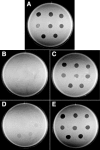
Effect of mutations in gldJ on bacteriophage resistance. Bacteriophages (5 μl of lysates containing approximately 6 × 10⁷ phage/ml) were spotted onto lawns of cells in CYE overlay agar containing 100 μg of erythromycin/ml. The plates were incubated at 25°C for 24 h to observe lysis. Bacteriophages were spotted in the following order from left to right: top row, φCj1, φCj13, and φCj23; middle row, φCj28, φCj29, and φCj42; bottom row, φCj48 and φCj54. (A) Wild-type F. johnsoniae with shuttle vector pCP11. (B) gldJ mutant UW102-55 with pCP11. (C) UW102-55 complemented with pMM313, which carries gldJ. (D) gldJ mutant UW102-48 with pCP11. (E) UW102-48 complemented with pMM313, which carries gldJ. The diameter of the petri dish is 9 cm.
FIG. 8.
FIG. 8.
Effect of mutation in gldJ on ability to utilize chitin. Approximately 4 × 10⁷ cells of wild-type F. johnsoniae with shuttle vector pCP11 (A), of the gldJ mutant UW102-48 with pCP11 (B), and of UW102-48 complemented with pMM313, which carries gldJ (C), were spotted on MYA-chitin medium containing 100 μg of erythromycin/ml and incubated for 4 days at 25°C.

References

    1. Agarwal, S., D. W. Hunnicutt, and M. J. McBride. 1997. Cloning and characterization of the Flavobacterium johnsoniae (Cytophaga johnsonae) gliding motility gene, gldA. Proc. Natl. Acad. Sci. USA 94:12139-12144. - PMC - PubMed
    1. Altschul, S. F., W. Gish, W. Miller, E. W. Myers, and D. J. Lipman. 1990. Basic local alignment search tool. J. Mol. Biol. 215:403-410. - PubMed
    1. Bayley, D. P., E. R. Rocha, and C. J. Smith. 2000. Analysis of cepA and other Bacteroides fragilis genes reveals a unique promoter structure. FEMS Microbiol. Lett. 193:149-154. - PubMed
    1. Beatson, P. J., and K. C. Marshall. 1994. A proposed helical mechanism for gliding motility in three gliding bacteria (order Cytophagales). Can. J. Microbiol. 40:173-183.
    1. Bhaya, D., N. R. Bianco, D. Bryant, and A. R. Grossman. 2000. Type IV pilus biogenesis and motility in the cyanobacterium Synechocystis sp. PCC6803. Mol. Microbiol. 37:941-951. - PubMed

Publication types

MeSH terms

Associated data

LinkOut - more resources