. 2005 Apr 4;33(6):1924-34.
doi: 10.1093/nar/gki339. Print 2005.

Regulation of Id2 expression by CCAAT/enhancer binding protein beta

Affiliations

Regulation of Id2 expression by CCAAT/enhancer binding protein beta

Kazuhiro Karaya et al. Nucleic Acids Res. .

Abstract

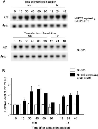
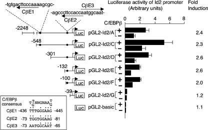
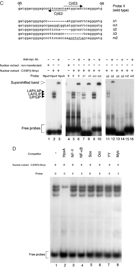
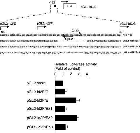
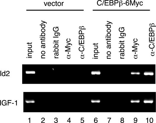
Mice deficient for Id2, a negative regulator of basic helix-loop-helix (bHLH) transcription factors, exhibit a defect in lactation due to impaired lobuloalveolar development during pregnancy, similar to the mice lacking the CCAAT enhancer binding protein (C/EBP) beta. Here, we show that Id2 is a direct target of C/EBPbeta. Translocation of C/EBPbeta into the nucleus, which was achieved by using a system utilizing the fusion protein between C/EBPbeta and the ligand-binding domain of the human estrogen receptor (C/EBPbeta-ERT), demonstrated the rapid induction of endogenous Id2 expression. In reporter assays, transactivation of the Id2 promoter by C/EBPbeta was observed and, among three potential C/EBPbeta binding sites found in the 2.3 kb Id2 promoter region, the most proximal element was responsible for the transactivation. Electrophoretic mobility shift assay (EMSA) identified this element as a core sequence to which C/EBPbeta binds. Chromatin immunoprecipitation (ChIP) furthermore confirmed the presence of C/EBPbeta in the Id2 promoter region. Northern blotting showed that Id2 expression in C/EBPbeta-deficient mammary glands was reduced at 10 days post coitus (d.p.c.), compared with that in wild-type mammary glands. Thus, our data demonstrate that Id2 is a direct target of C/EBPbeta and provide insight into molecular mechanisms underlying mammary gland development during pregnancy.


Figures

Figure 1
Figure 1
Induction of endogenous Id2 expression by the C/EBPβ-ERT fusion protein. A permanent cell line expressing C/EBPβ-ERT was established with NIH3T3 cells and treated with 1 μM 4HT to induce the translocation of the C/EBPβ-ERT fusion protein from the cytosol. RNA was extracted from the cells at various times and subjected to northern blot analysis. Representative results are shown in (A). The probes used are indicated on the left of each panel. Relative Id2 expression is shown in (B). Actb expression was used as an internal control. Each Id2 mRNA level is expressed as fold increase, compared to that in cells incubated without 4HT. White and black bars indicate relative Id2 mRNA levels in the parental NIH3T3 cells and NIH3T3 cells stably expressing C/EBPβ-ERT, respectively. Results are shown as the mean and standard error values of the mean (n = 3).
Figure 2
Figure 2
Transactivation of the Id2 promoter by C/EBPβ. In the Id2 promoter region spanning positions −2248 to +84 from the transcription initiation site, there are three potential C/EBPβ binding sites, named CβE1 (−445 to −436), CβE2 (−81 to −73) and CβE3 (−73 to −65), as indicated by arrows in the schematic representation of pGL2-Id2/A. The numbers shown on the left of the respective reporter constructs indicate the positions from the transcription initiation site. The sequence comparison of these sites with the consensus binding site (8), TT/GNNGNAAT/G, is shown in the inset. In the reporter assay, each of the reporter plasmids, which contained various lengths of the Id2 promoter, was co-transfected into NIH3T3 cells with pPGK-renilla, together with pMSV-C/EBPβ or pMSV-mock, as indicated by + and −, respectively, in the middle, and luciferase activities were determined after 48 h. Luciferase activities of the respective reporter constructs were determined by normalizing by the respective renilla luciferase activity. Fold induction of luciferase activity by C/EBPβ is indicated on the right. The mean and standard error of the mean values of three independent experiments are shown.
Figure 3
Figure 3
EMSA for C/EBPβ with the Id2 promoter. EMSA was performed with 32P-labeled probes and nuclear extracts obtained from NIH3T3 cells transiently transfected with pC/EBPβ-6Myc or parental NIH3T3 cells. Oligonucleotide HpxA was used as a positive control for C/EBPβ binding. DNA–protein complexes were separated on a 6% polyacrylamide gel, and the gel was dried and exposed to X-ray film. Three retarded bands derived from dimers of C/EBPβ isoforms are indicated by filled arrowheads (LAP/LAP, LAP/LIP and LIP/LIP). The supershifted band formed with anti-Myc Ab is indicated by an open arrowhead. (A) On the top, probes used in EMSA are aligned with their positions in the Id2 promoter sequence. Arrows indicate the positions of CβE2 and CβE3. The probes used were HpxA (lanes 1–3), probe I (lanes 4–6), probe II (lanes 7–9), probe III (lanes 10–12) and probe IV (lanes 13–15). Nuclear extracts were prepared from NIH3T3 cells transfected with pC/EBPβ-6Myc (lanes 2, 3, 5, 6, 8, 9, 11, 12, 14, 15) or the parental NIH3T3 cells (lanes 1, 4, 7, 10, 13). For the detection of a supershifted band, anti-Myc Ab was included in the incubation mixture (lanes 3, 6, 9, 12, 15). In lanes 16 and 17, a 200-fold molar excess of the HpxA oligonucleotide was added as cold competitor. (B) Expression of C/EBPβ isoforms in NIH3T3 cells. Left and right panels show the results with anti-C/EBPβ and anti-Myc Abs, respectively. Origins of nuclear extracts are shown on the top. Positions of the respective C/EBPβ isoforms are indicated by arrowheads (untagged-C/EBPβ) and arrows (6Myc-tagged-C/EBPβ) on the right of each panel. Positions of size markers are indicated on the left of each panel. (C) Mutant oligonucleotides designed based on probe II were used in EMSA. The positions of CβE2 and CβE3 are indicated by arrows. Probes Δ1 and Δ3 are CβE2 and CβE3 deletion mutants of probe II, respectively. Deleted regions are indicated by dashes. Base-substituted regions are underlined. The probes used were HpxA (lanes 1–3), probe II (lanes 4–6), Δ1 (lanes 7 and 8), m1 (lanes 9 and 10), Δ2 (lanes 11 and 12), Δ3 (lanes 13 and 14) and m2 (lanes 15 and 16). Nuclear extracts were prepared from NIH3T3 cells transfected with pC/EBPβ-6Myc (lanes 2, 3, 5–16) or the parental NIH3T3 cells (lanes 1 and 4). For the detection of a supershifted band, anti-Myc Ab was included in the incubation mixture (lanes 3, 6, 8, 12, 14 and 16). (D) 32P-labeled probe II was incubated with nuclear extracts containing C/EBPβ-6Myc, together with 200-fold molar excess of the cold competitors that contain the binding sites of the transcription factors as indicated on the top (see Materials and Methods).
Figure 3
Figure 3
EMSA for C/EBPβ with the Id2 promoter. EMSA was performed with 32P-labeled probes and nuclear extracts obtained from NIH3T3 cells transiently transfected with pC/EBPβ-6Myc or parental NIH3T3 cells. Oligonucleotide HpxA was used as a positive control for C/EBPβ binding. DNA–protein complexes were separated on a 6% polyacrylamide gel, and the gel was dried and exposed to X-ray film. Three retarded bands derived from dimers of C/EBPβ isoforms are indicated by filled arrowheads (LAP/LAP, LAP/LIP and LIP/LIP). The supershifted band formed with anti-Myc Ab is indicated by an open arrowhead. (A) On the top, probes used in EMSA are aligned with their positions in the Id2 promoter sequence. Arrows indicate the positions of CβE2 and CβE3. The probes used were HpxA (lanes 1–3), probe I (lanes 4–6), probe II (lanes 7–9), probe III (lanes 10–12) and probe IV (lanes 13–15). Nuclear extracts were prepared from NIH3T3 cells transfected with pC/EBPβ-6Myc (lanes 2, 3, 5, 6, 8, 9, 11, 12, 14, 15) or the parental NIH3T3 cells (lanes 1, 4, 7, 10, 13). For the detection of a supershifted band, anti-Myc Ab was included in the incubation mixture (lanes 3, 6, 9, 12, 15). In lanes 16 and 17, a 200-fold molar excess of the HpxA oligonucleotide was added as cold competitor. (B) Expression of C/EBPβ isoforms in NIH3T3 cells. Left and right panels show the results with anti-C/EBPβ and anti-Myc Abs, respectively. Origins of nuclear extracts are shown on the top. Positions of the respective C/EBPβ isoforms are indicated by arrowheads (untagged-C/EBPβ) and arrows (6Myc-tagged-C/EBPβ) on the right of each panel. Positions of size markers are indicated on the left of each panel. (C) Mutant oligonucleotides designed based on probe II were used in EMSA. The positions of CβE2 and CβE3 are indicated by arrows. Probes Δ1 and Δ3 are CβE2 and CβE3 deletion mutants of probe II, respectively. Deleted regions are indicated by dashes. Base-substituted regions are underlined. The probes used were HpxA (lanes 1–3), probe II (lanes 4–6), Δ1 (lanes 7 and 8), m1 (lanes 9 and 10), Δ2 (lanes 11 and 12), Δ3 (lanes 13 and 14) and m2 (lanes 15 and 16). Nuclear extracts were prepared from NIH3T3 cells transfected with pC/EBPβ-6Myc (lanes 2, 3, 5–16) or the parental NIH3T3 cells (lanes 1 and 4). For the detection of a supershifted band, anti-Myc Ab was included in the incubation mixture (lanes 3, 6, 8, 12, 14 and 16). (D) 32P-labeled probe II was incubated with nuclear extracts containing C/EBPβ-6Myc, together with 200-fold molar excess of the cold competitors that contain the binding sites of the transcription factors as indicated on the top (see Materials and Methods).
Figure 4
Figure 4
Transactivation of the Id2 promoter by C/EBPβ is via CβE3. Mutant reporter plasmids were constructed based on pGL2-Id2/E. pGL2-Id2/EΔ1 lacks the region containing both CβE2 and CβE3. Core sequences of CβE2 and CβE3 are deleted in pGL2-Id2/EΔ2 and pGL2-Id2/EΔ3, respectively. Deleted regions are indicated by dashes. NIH3T3 cells were co-transfected with reporter plasmids and an internal control vector, pGL-renilla, together with pMSV-C/EBPβ or pMSV-mock. Luciferase activity was determined as in the experiments shown in Figure 2. Transactivation of the respective reporter plasmids by C/EBPβ is presented as fold induction of transactivation of pGL2-basic by C/EBPβ. The mean and standard errors of the mean values from three separate assays are shown in the histograms.
Figure 5
Figure 5
Binding of C/EBPβ to the Id2 promoter in vivo. The ChIP assay was performed using NIH3T3 cells transiently transfected with empty vector (lanes 1–5) or pC/EBPβ-6Myc (lanes 6–10). The chromatin-associated DNA was incubated without Ab (lanes 2 and 7) or with normal rabbit IgG (lanes 3 and 8), anti-Myc Ab (lanes 4 and 9), or anti-C/EBPβ Ab (lanes 5 and 10). An aliquot (2.5%) of the total chromatin DNA was used for input (lanes 1 and 6). Immunoprecipitates were subjected to PCR with a primer-pair specific to the Id2 promoter that amplified a 311 bp fragment (upper panel). As a positive control, PCR was carried out with a primer-pair specific to the Igf-1 promoter, which amplified a 287 bp DNA fragment (lower panel). After 32 cycles of amplification, PCR products were electrophoresed through a 3% agarose gel and visualized by ethidium bromide staining.
Figure 6
Figure 6
Id2 expression in mammary glands deficient for C/EBPβ. Epithelial cells of wild-type or C/EBPβ-null mammary glands were transplanted into precleared mammary glands of female Rag2-null mice. After 7 weeks, reconstituted mammary glands of non-pregnant and pregnant (10 d.p.c.) mice were subjected to whole mount analysis and RNA purification. (A) Morphology of reconstituted mammary glands in the non-pregnant and pregnant mice. Reconstituted mammary glands were dissected out, mounted on a slide glass and stained with carmine. The genotype of a donor and the pregnant state of a recipient are indicated on the left and the top, respectively. Both wild-type and C/EBPβ-null mammary epithelial cells reconstituted the mammary gland of a recipient successfully and no significant difference was observed in the non-pregnant state. In the mammary gland at 10 d.p.c., disturbed epithelial proliferation is evident in the gland reconstituted with C/EBPβ-null mammary epithelial cells. (B) Northern blot analysis of Id2 mRNA expression. RNA was purified from reconstituted mammary glands. Genotypes of transplanted mammary epithelial cells are indicated on the top. The lower panel shows the representative data. The probes used are indicated on the left of the lower panels. The upper panel shows the relative Id2 expression normalized by Cyk18 expression. The mean and standard error of the mean values obtained from three independent samples are shown in the histograms.

Similar articles

Cited by

References

    1. Neville M.C., Daniel C.W. The Mammary gland: Development, Regulation, and Function. New York, NY: Plenum Press; 1987.
    1. Hennighausen L., Robinson G.W. Signaling pathways in mammary gland development. Dev. Cell. 2001;1:467–475. - PubMed
    1. Ruzinova M.B., Benezra R. Id proteins in development, cell cycle and cancer. Trends Cell Biol. 2003;13:410–418. - PubMed
    1. Mori S., Nishikawa S., Yokota Y. Lactation defect in mice lacking the helix–loop–helix inhibitor Id2. EMBO J. 2000;19:5772–5781. - PMC - PubMed
    1. Takiguchi M. The C/EBP family of transcription factors in the liver and other organs. Int. J. Exp. Pathol. 1998;79:369–391. - PMC - PubMed

Publication types

MeSH terms

Substances