

Phosphoinositide3-kinase regulates actin polymerization during delayed phagocytosis of Helicobacter pylori
- PMID: 15809290
- PMCID: PMC1868428
- DOI: 10.1189/jlb.0205091
Phosphoinositide3-kinase regulates actin polymerization during delayed phagocytosis of Helicobacter pylori
Abstract
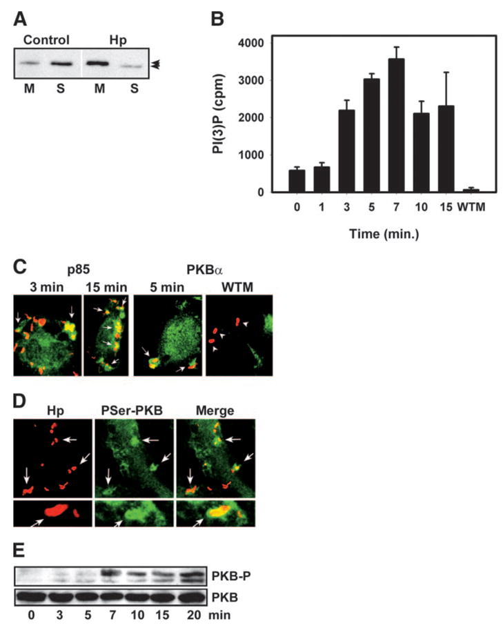
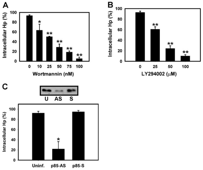
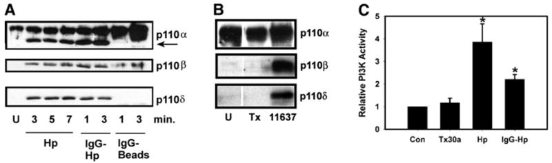
We have shown previously that ulcerogenic (type I) strains of Helicobacter pylori (Hp) retard their entry into macrophages. However, the signaling pathways that regulate Hp phagocytosis are largely undefined. We show here that Hp strongly activated class IA phosphoinositide3-kinases (PI3Ks) in macrophages, coincident with phagocytosis, and endogenous p85 and active protein kinase Balpha accumulated on forming phagosomes. PI3K inhibitors, wortmannin and LY294002, inhibited phagocytosis of Hp in a dose-dependent manner, and blockade of engulfment correlated directly with loss of 3'-phosphoinositides in the membrane subjacent to attached bacteria. During uptake of large immunoglobulin G (IgG)-coated particles, PI3Ks regulate pseudopod extension and phagosome closure. In marked contrast, we show here that 3'-phosphoinositides regulated actin polymerization at sites of Hp uptake. Moreover, Hp and IgG beads activated distinct PI3K isoforms. Phagosomes containing IgG-coated particles accumulated 3'-phosphatase and tensin homologue deleted on chromosome 10 and Src homology 2 domain-containing inositol 5'-phosphatase, yet Hp phagosomes did not. Finally, rapid uptake of IgG-opsonized Hp or a less-virulent type II Hp was PI3K-independent. We conclude that Hp and IgG beads are ingested by distinct mechanisms and that PI3Ks regulate the actin cytoskeleton during slow phagocytosis of ulcerogenic Hp.
Figures


Similar articles
-
Atypical protein kinase C-zeta is essential for delayed phagocytosis of Helicobacter pylori.Curr Biol. 2002 Oct 15;12(20):1762-6. doi: 10.1016/s0960-9822(02)01216-2. Curr Biol. 2002. PMID: 12401171
-
Critical role for scaffolding adapter Gab2 in Fc gamma R-mediated phagocytosis.J Cell Biol. 2003 Jun 23;161(6):1151-61. doi: 10.1083/jcb.200212158. J Cell Biol. 2003. PMID: 12821647 Free PMC article.
-
Restricted accumulation of phosphatidylinositol 3-kinase products in a plasmalemmal subdomain during Fc gamma receptor-mediated phagocytosis.J Cell Biol. 2001 Jun 25;153(7):1369-80. doi: 10.1083/jcb.153.7.1369. J Cell Biol. 2001. PMID: 11425868 Free PMC article.
-
Phosphatases as small-molecule targets: inhibiting the endogenous inhibitors of kinases.Biochem Soc Trans. 2004 Apr;32(Pt 2):348-9. doi: 10.1042/bst0320348. Biochem Soc Trans. 2004. PMID: 15046606 Review.
-
Phosphatidylinositol Kinases and Phosphatases in Entamoeba histolytica.Front Cell Infect Microbiol. 2019 Jun 6;9:150. doi: 10.3389/fcimb.2019.00150. eCollection 2019. Front Cell Infect Microbiol. 2019. PMID: 31245297 Free PMC article. Review.
Cited by
-
Revaprazan, a novel acid pump antagonist, exerts anti-inflammatory action against Helicobacter pylori-induced COX-2 expression by inactivating Akt signaling.J Clin Biochem Nutr. 2012 Sep;51(2):77-83. doi: 10.3164/jcbn.11-94. Epub 2012 Jul 20. J Clin Biochem Nutr. 2012. PMID: 22962522 Free PMC article.
-
Helicobacter pylori promotes angiogenesis depending on Wnt/beta-catenin-mediated vascular endothelial growth factor via the cyclooxygenase-2 pathway in gastric cancer.BMC Cancer. 2016 May 19;16:321. doi: 10.1186/s12885-016-2351-9. BMC Cancer. 2016. PMID: 27198692 Free PMC article.
-
Investigating the Role of Helicobacter pylori PriA Protein.Helicobacter. 2016 Aug;21(4):295-304. doi: 10.1111/hel.12283. Epub 2016 Jan 28. Helicobacter. 2016. PMID: 26817518 Free PMC article.
-
CD300ld promotes neutrophil bacterial phagocytosis in sepsis.J Leukoc Biol. 2025 May 7;117(5):qiaf063. doi: 10.1093/jleuko/qiaf063. J Leukoc Biol. 2025. PMID: 40376837
-
Dysregulated macrophage immunity in Helicobacter pylori infection: unveiling mechanistic insights and therapeutic implications.Front Immunol. 2025 Aug 4;16:1636768. doi: 10.3389/fimmu.2025.1636768. eCollection 2025. Front Immunol. 2025. PMID: 40831563 Free PMC article. Review.
References
-
- Covacci A, Telford JL, Del Giudice G, Parsonnet J, Rappuoli R. Helicobacter pylori virulence and genetic geography. Science. 1999;284:1328 –1333. - PubMed
-
- Xiang Z, Censini S, Bayeli PF, Telford JL, Figura N, Rappuoli R, Covacci A. Analysis of expression of CagA and VacA virulence factors in 43 strains of Helicobacter pylori reveals that clinical isolates can be divided into two major types and that CagA is not necessary for expression of the vacuolating cytotoxin. Infect Immun. 1995;63:94 –98. - PMC - PubMed
-
- Andersen LP, Blom J, Nielsen H. Survival and ultrastructural changes of Helicobacter pylori after phagocytosis by human polymorpho-nuclear phagocytes and monocytes. APMIS. 1993;101:61–72. - PubMed
-
- Engstrand L, Graham D, Scheynius A, Genta RM, ElZaatari F. Is the sanctuary where Helicobacter pylori avoids antibacterial treatment intracellular? Am J Clin Pathol. 1997;108:504 –509. - PubMed
Publication types
MeSH terms
Substances
Grants and funding
LinkOut - more resources
Full Text Sources
Medical
Research Materials
Miscellaneous
