Comparative Study
. 2005 Apr 4;201(7):1089-99.
doi: 10.1084/jem.20041896.

VEGF-A induces tumor and sentinel lymph node lymphangiogenesis and promotes lymphatic metastasis

Affiliations
Comparative Study

VEGF-A induces tumor and sentinel lymph node lymphangiogenesis and promotes lymphatic metastasis

Satoshi Hirakawa et al. J Exp Med. .

Abstract

The mechanisms of tumor metastasis to the sentinel lymph nodes are poorly understood. Vascular endothelial growth factor (VEGF)-A plays a principle role in tumor progression and angiogenesis; however, its role in tumor-associated lymphangiogenesis and lymphatic metastasis has remained unclear. We created transgenic mice that overexpress VEGF-A and green fluorescent protein specifically in the skin, and subjected them to a standard chemically-induced skin carcinogenesis regimen. We found that VEGF-A not only strongly promotes multistep skin carcinogenesis, but also induces active proliferation of VEGF receptor-2-expressing tumor-associated lymphatic vessels as well as tumor metastasis to the sentinel and distant lymph nodes. The lymphangiogenic activity of VEGF-A-expressing tumor cells was maintained within metastasis-containing lymph nodes. The most surprising finding of our study was that even before metastasizing, VEGF-A-overexpressing primary tumors induced sentinel lymph node lymphangiogenesis. This suggests that primary tumors might begin preparing their future metastatic site by producing lymphangiogenic factors that mediate their efficient transport to sentinel lymph nodes. This newly identified mechanism of inducing lymph node lymphangiogenesis likely contributes to tumor metastasis, and therefore, represents a new therapeutic target for advanced cancer and/or for the prevention of metastasis.


Figures

Figure 1.
Figure 1.
K14/GFP transgenic mice provide a reliable new model for the detection of skin cancer metastases. (A) Schematic representation of the K14-GFP transgenic construct. A 720-bp enhanced GFP (EGFP) cDNA fragment was ligated to the BamHI restriction site of the keratin 14 promoter cassette. (B) Targeted expression of the K14/GFP transgene in the basal epidermal keratinocyte layer and in outer root sheath keratinocytes of hair follicles was confirmed by fluorescence microscopy (green). (C and D) Induction of epidermal hyperplasia by topical application of PMA resulted in strong GFP expression throughout the epidermis. Nuclei are labeled blue (Hoechst stain). (E and F) High levels of GFP expression (green) were maintained in tumor cells of papillomas (E) and of SCC (F) induced by the chemical skin carcinogenesis regimen. Immunostains for the panvascular marker CD31 (E; red) revealed tumor-associated angiogenesis, whereas immunostains for the lymphatic-specific marker LYVE-1 (F; red; arrowheads) demonstrated lymphangiogenesis in close association with tumor cells at the advancing tumor edge. (G and H) Metastatic tumor cells in sentinel lymph nodes of K14/GFP transgenic mice maintained high levels of GFP expression, enabling simple detection of lymph node metastasis by fluorescence microscopy (G, green) and quantification of GFP-positive and CD45-negative metastatic tumor cells (66.2% of all cells in this representative case) by FACS analysis of single cell suspensions (H). Bars, 100 μm (B–F), 500 μm (G).
Figure 2.
Figure 2.
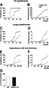
Accelerated and increased skin carcinogenesis in VEGF-A transgenic mice. (A) Accelerated development of skin papillomas in VEGF-A transgenic mice (n = 34; filled squares), as compared with wild-type mice (n = 31; open circles). Incidence is expressed as the percentage of mice with detectable papillomas (>1 mm) during the 20 wk of topical PMA application. (B) Significant increase in frequency of papilloma formation in VEGF-A transgenic mice, expressed as the average number of papillomas per mouse. P < 0.01 at wk 6, P < 0.001 from wk 7 to 20. (C) Accelerated development of large papillomas (>3 mm) in VEGF-A transgenic mice. (D) Increased frequency of large papillomas in VEGF-A transgenic mice. P < 0.01 from wk 11 to 13; P < 0.001 from wk 14 to 20. (E) Increased incidence of SCC in VEGF-A transgenic mice. (F) Increased number of SCC per mouse in VEGF-A transgenic mice. P < 0.01 from wk 19 to 22; P < 0.001 after 23 wk. (G) Comparable ratio of malignant conversion of large papillomas into SCC in VEGF-A transgenic and wild-type mice.
Figure 3.
Figure 3.
Enhanced tumor angiogenesis and lymphangiogenesis in VEGF-A transgenic mice. Immunofluorescence analysis with antibodies against CD31 (green) and LYVE-1 (red) of PMA-treated skin (A and B), early papillomas (C and D) and SCC (E and F) of wild-type (A,C,E) and VEGF-A transgenic mice (B,D,F) demonstrate highly increased vascularization of papillomas and SCC in both genotypes, as compared with PMA-treated skin. Tumor angiogenesis and lymphangiogenesis were more prominent in VEGF-A transgenic mice (D and F) than in wild-type mice (C and E). Note enlargement of blood vessels (green) and lymphatic vessels (red) in VEGF-A transgenic mice. (G and H) Double-immunofluorescence analysis of SCC for the proliferation marker BrdU (green; arrowheads) and the lymphatic marker LYVE-1 (red) revealed numerous proliferating lymphatic endothelial cells in VEGF-A transgenic mice (H), as compared with only occasional proliferating lymphatic endothelial cells observed in wild-type mice (G). This indicates that VEGF-A promotes tumor lymphangiogenesis. Nuclei are labeled blue (Hoechst stain). Scale bars = 100 μm. (I–N) Computer- assisted morphometric analysis of normal cutaneous and of tumor-associated lymphatic and blood vessels. (I) Significant increase of the relative area occupied by blood vessels in the peritumoral area (Peri SCC) as well as within SCC (Intra SCC), in VEGF-A transgenic mice (filled bars), as compared with wild-type mice (open bars). (J) The average blood vessel size was increased significantly in the intratumoral and the peritumoral areas of SCC in VEGF-A transgenic mice, as compared with wild-types, whereas the blood vessel density only was increased in the intratumoral areas (K). (L) Significant increase of the relative area occupied by lymphatic vessels in VEGF-A transgenic mice throughout all stages of skin carcinogenesis. (M) Significantly increased lymphatic vessel size in the peritumoral area of SCC, but not in the intratumoral area of VEGF-A transgenic mice. (N) No significant differences were found in tumor-associated lymphatic vessel density between the two genotypes. Data are expressed as mean ± SEM. *P < 0.05; **P < 0.01; ***P < 0.001.
Figure 4.
Figure 4.
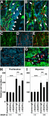
Formation of tumor-associated lymphatic vessels in VEGF-A transgenic mice. (A and B) Double immunofluorescence stains for CD31 (green) and LYVE-1 (red) reveal initial invasion of angiogenic blood vessels (green; arrowheads), but not of lymphatic vessels (red) into benign papillomas (A), whereas intratumoral blood vessels often are accompanied by newly formed lymphatic vessels (arrows) in primary cutaneous SCC (B) of VEGF-A transgenic mice. (C–E) Intratumoral LYVE-1–positive lymphatic vessels (D; red) in SCC express VEGFR-2 (C; green; E = merged image; orange/yellow). Green vessels in panel E are LYVE-1-negative blood vessels. (F) All tumor-associated LYVE-1–positive vessels (red) also express the lymphatic-specific transcription factor Prox1 (green). (G) GFP-expressing SCC cells (arrowheads) were found attached to LYVE-1–positive lymphatic vessels (red; asterisks). Nuclei are labeled blue (Hoechst stain). Scale bars = 200 μm (A and B); 100 μm (C–F); 50 μm (G). (H) VEGF-A induced LEC proliferation is inhibited significantly by antibody blockade of VEGFR-2, but not of VEGFR-3. (I) Blockade of VEGFR-2, but not of VEGFR-3, inhibits VEGF-A–induced LEC migration. Data are expressed as mean ± SD (n = 5 per group) and are representative for three independent experiments. **P < 0.01; ***P < 0.001.
Figure 5.
Figure 5.
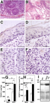
Expression of VEGF-A and VEGF-C by SCC of VEGF-A transgenic mice. (A) Representative histologic image (hematoxylin-eosin stain) of an SCC from a wild-type mouse shows nests of tumor cells surrounded by compact stroma that contain small vessels. (B) SCC observed in VEGF-A transgenic mice were characterized by tissue edema and vessel enlargement. Scale bars = 200 μm (A and B). (C–F) In situ hybridization for VEGF-A and VEGF-C mRNA expression in SCC of VEGF-A transgenic mice. Normal epidermal keratinocytes of the overlying epidermis (C) and SCC tumor cells (E) express high levels of VEGF-A mRNA, whereas stromal cells show little or no expression. Occasionally, low levels of focal VEGF-C expression were observed in the overlying epidermis (D) and in SCC cells (F). Scale bars = 50 μm (C–F). (G) ELISA analysis of VEGF-A expression in skin and tumor lysates (n = 5 per group) revealed significantly increased levels of VEGF-A protein in SCC of both genotypes, as compared with normal skin. VEGF-A levels were higher in VEGF-A transgenic mice than in wild-type mice. (H) Quantitative real-time TaqMan RT-PCR demonstrated increased levels of VEGF-C mRNA expression in the skin and in SCC of VEGF-A transgenic mice compared with wild-type mice. VEGF-C expression was increased significantly in SCC of VEGF-A transgenic mice, as compared with normal skin. *P < 0.05; **P < 0.01; ***P < 0.001. (I) Immunoprecipitation of SCC lysates reveals increased levels of the 58-kD VEGF-C propeptide in VEGF-A transgenic tumors, as compared with wild-type tumors. No major differences in the proteolytic processing of VEGF-C protein were found between the two genotypes. Results are representative for five independent experiments.
Figure 6.
Figure 6.
Increased metastasis to the sentinel lymph nodes in VEGF-A transgenic mice. (A and C) Representative flow cytometry analysis of GFP-expressing, CD45-negative cells in primary SCC and in sentinel lymph node (LN). Whereas no GFP-positive cells were found in nontransgenic control mice at 8 wk after the first detection of cutaneous SCC, 53.5% of all SCC-derived cells in K14/GFP transgenic mice and 67.9% in K14/GFP/VEGF-A transgenic mice were GFP-positive (A). In contrast, the percentage of GFP-expressing tumor cells was significantly higher in metastases to the sentinel lymph nodes of K14/GFP/VEGF-A transgenic mice (50.2%) than of K14/GFP transgenic mice (4.9%; C). (B and D) Statistical analysis revealed a significant increase of GFP-positive tumor cells in the sentinel lymph node metastases of VEGF-A/GFP double transgenic mice, compared with GFP transgenic mice (D), whereas no significant differences were found in the primary lesions (B). Data are expressed as mean ± SEM (n = 5 per group). ***P < 0.001.
Figure 7.
Figure 7.
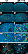
Increased lymph node lymphangiogenesis in VEGF-A transgenic mice. Double immunofluorescence stains of lymph nodes of nontumor-bearing mice demonstrate a comparable pattern of CD31-positive postcapillary high endothelial venules (green) and LYVE-1–positive sinusoids (red) in wild-type (WT) mice (A) and in VEGF-A transgenic (VEGF-TG) mice (B). Nonmetastatic sentinel lymph nodes of SCC-bearing VEGF-A transgenic mice have increased numbers of enlarged LYVE-1–positive sinusoids (red; D), as compared with wild-type mice (C). Increased numbers of enlarged LYVE-1-positive lymphatic vessels (red) and of CD31-positive blood vessels (green) were found in the metastatic sentinel lymph nodes of VEGF-A transgenic mice (F), as compared with wild-type mice (E). (G and H) Higher magnification of panels E and F, respectively. The tumor-associated LYVE-1–positive vessels (red) in VEGF-A transgenic mice had high levels of BrdU staining in lymphatic endothelial cells (I; green), indicating active lymphatic proliferation (arrowheads) within the sentinel lymph node. These cells also expressed Prox1 (J; green). Blood vessels (J; asterisks) do not express LYVE-1 or Prox1. ‘Met’ indicates a metastasizing cutaneous SCC. Nuclei are labeled blue (Hoechst stain). Scale bars = 100 μm (A–D and I); 200 μm (E–H); 50 μm (J).

Similar articles

Cited by

References

    1. Folkman, J. 1992. The role of angiogenesis in tumor growth. Semin. Cancer Biol. 3:65–71. - PubMed
    1. Ferrara, N., H.P. Gerber, and J. LeCouter. 2003. The biology of VEGF and its receptors. Nat. Med. 9:669–676. - PubMed
    1. Skobe, M., T. Hawighorst, D.G. Jackson, R. Prevo, L. Janes, P. Velasco, L. Riccardi, K. Alitalo, K. Claffey, and M. Detmar. 2001. Induction of tumor lymphangiogenesis by VEGF-C promotes breast cancer metastasis. Nat. Med. 7:192–198. - PubMed
    1. Stacker, S.A., C. Caesar, M.E. Baldwin, G.E. Thornton, R.A. Williams, R. Prevo, D.G. Jackson, S. Nishikawa, H. Kubo, and M.G. Achen. 2001. VEGF-D promotes the metastatic spread of tumor cells via the lymphatics. Nat. Med. 7:186–191. - PubMed
    1. Mandriota, S.J., L. Jussila, M. Jeltsch, A. Compagni, D. Baetens, R. Prevo, S. Banerji, J. Huarte, R. Montesano, D.G. Jackson, et al. 2001. Vascular endothelial growth factor-C-mediated lymphangiogenesis promotes tumour metastasis. EMBO J. 20:672–682. - PMC - PubMed

Publication types

MeSH terms

Substances