. 2005 Apr 12;102(15):5588-93.
doi: 10.1073/pnas.0501703102. Epub 2005 Apr 4.

The contribution of GABA to glutamate/glutamine cycling and energy metabolism in the rat cortex in vivo

Affiliations

The contribution of GABA to glutamate/glutamine cycling and energy metabolism in the rat cortex in vivo

Anant B Patel et al. Proc Natl Acad Sci U S A. .

Abstract

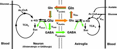
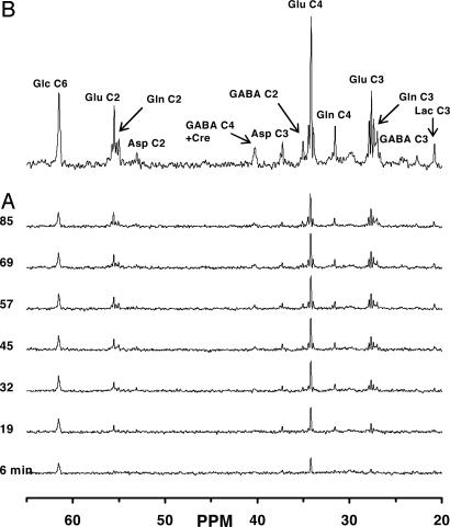
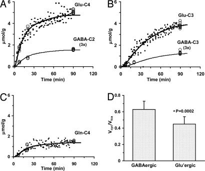
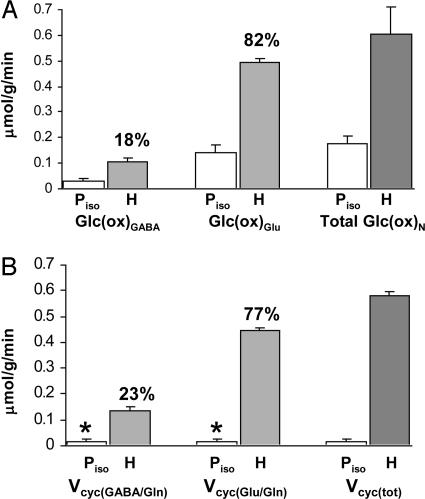
Previous studies have shown that the glutamate/glutamine (Glu/Gln) neurotransmitter cycle and neuronal glucose oxidation are proportional (1:1), with increasing neuronal activity above isoelectricity. GABA, a product of Glu metabolism, is synthesized from astroglial Gln and contributes to total Glu/Gln neurotransmitter cycling, although the fraction contributed by GABA is unknown. In the present study, we used (13)C NMR spectroscopy together with i.v. infusions of [1,6-(13)C(2)]glucose and [2-(13)C]acetate to separately determine rates of Glu/Gln and GABA/Gln cycling and their respective tricarboxylic acid cycles in the rat cortex under conditions of halothane anesthesia and pentobarbital-induced isoelectricity. Under 1% halothane anesthesia, GABA/Gln cycle flux comprised 23% of total (Glu plus GABA) neurotransmitter cycling and 18% of total neuronal tricarboxylic acid cycle flux. In isoelectric cortex, glucose oxidation was reduced >3-fold in glutamatergic and GABAergic neurons, and neurotransmitter cycling was below detection. Hence, in both cell types, the primary energetic costs are associated with neurotransmission, which increase together as cortical activity is increased. The contribution of GABAergic neurons and inhibition to cortical energy metabolism has broad implications for the interpretation of functional imaging signals.


Figures

Fig. 1.
Fig. 1.
Schematic depiction of Glu/Gln and GABA/Gln cycling between glutamatergic (orange) and GABAergic (green) neurons and astroglia relevant to 13C NMR experiments using [1-13C]glucose or [2-13C]acetate as tracer. Metabolism of [1-13C]glucose through glycolysis and the TCA cycle labels neuronal Glu-C4 and GABA-C2 with label transfer to Gln-C4 by neurotransmitter cycling, Vcyc(tot) = VGlu/Gln + VGABA/Gln. [2-13C]acetate metabolism in astroglia labels Gln-C4 directly with label transfer to neuronal Glu-C4 and GABA-C2 by neurotransmitter cycling, Vcyc(tot). VGAD, rate of GABA synthesis via Glu decarboxylase; Vshunt, rate of neuronal GABA catabolism; VGln, rate of Gln synthesis; and VPC, rate of pyruvate carboxylation.
Fig. 2.
Fig. 2.
Spatially localized 13C-[1H]NMR spectrum of metabolites in the intact rat brain after infusion of [1,6-13C2]glucose and acetate under halothane. Time courses of 13C-labeling of metabolites (A) and averaged spectrum over the last 30 min (60–90 min) of the 1.5-h [1,6-13C2]glucose and acetate infusion (B). Peak label definitions: Asp, aspartate; Lac, lactate; Glc, glucose.
Fig. 3.
Fig. 3.
Fit of the metabolic model to experimental data. The in vivo time course of 13C labeling of Glu-C4 (A), Glu-C3 (B), and Gln-C4 (C) from [1,6-13C2]glucose is depicted for all animals (data points). Ex vivo measurements at selected time points are depicted by open circles in (A) Glu-C4 and GABA-C2, (B) Glu-C3 and GABA-C3, and (C) Gln-C4. The best fit of the mathematical model to the experimental data is shown by the smooth curves. (D) Values of the ratio, Vcyc/VTCA, in glutamatergic and GABAergic neurons calculated from steady-state fractional enrichments of Glu-C4, Gln-C4, and GABA-C2 during infusion of [2-13C]acetate and glucose (Table 1) used as constraints in the data fitting shown in A–C.
Fig. 4.
Fig. 4.
Contribution of GABAergic and glutamatergic neurons to neuronal glucose oxidation and neurotransmitter cycling. (A) Rates of glucose oxidation, Glc(ox), for GABAergic and glutamatergic neurons and their sum, total Glc(ox)N. (B) Rates of GABA/Gln and Glu/Gln cycling and their sum, Vcyc(tot). Piso, pentobarbital-induced isoelectricity; H, halothane/N20 anesthesia. *, maximum possible value for Piso condition given by the measured value of Vcyc(tot) for isoelectricity. Percentages denote the contributions of GABAergic and glutamatergic neurons to the total fluxes given.

References

    1. Peters, A. & Jones, E. G. (1984) Cerebral Cortex: Cellular Components of the Cerebral Cortex (Plenum, New York).
    1. Beaulieu, C. & Colonnier, M. (1985) J. Comp. Neurol. 231, 180–189. - PubMed
    1. Sibson, N. R., Dhankhar, A., Mason, G. F., Behar, K. L., Rothman, D. L. & Shulman, R. G. (1997) Proc. Natl. Acad. Sci. USA 94, 2699–2704. - PMC - PubMed
    1. Sibson, N. R., Dhankhar, A., Mason, G. F., Rothman, D. L., Behar, K. L. & Shulman, R. G. (1998) Proc. Natl. Acad. Sci. USA 95, 316–321. - PMC - PubMed
    1. Sibson, N. R., Mason, G. F., Shen, J., Cline, G. W., Herskovits, A. Z., Wall, J. E., Behar, K. L., Rothman, D. L. & Shulman, R. G. (2001) J. Neurochem. 76, 975–989. - PubMed

Publication types

MeSH terms

LinkOut - more resources