. 2005 Apr 19;102(16):5856-61.
doi: 10.1073/pnas.0501634102. Epub 2005 Apr 4.

The CNS role of Toll-like receptor 4 in innate neuroimmunity and painful neuropathy

Affiliations

The CNS role of Toll-like receptor 4 in innate neuroimmunity and painful neuropathy

Flobert Y Tanga et al. Proc Natl Acad Sci U S A. .

Abstract

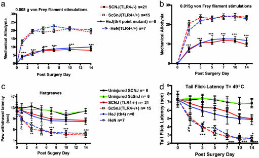
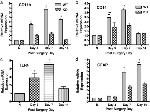
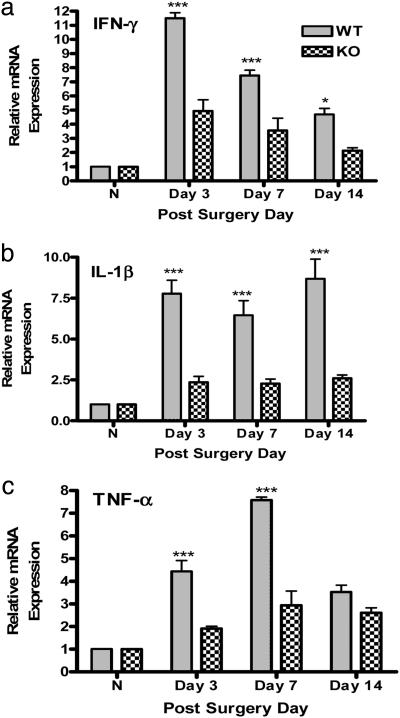
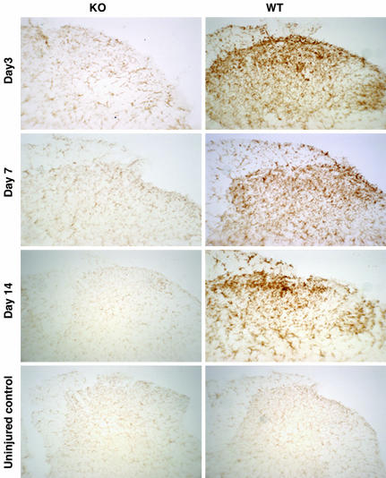
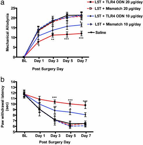
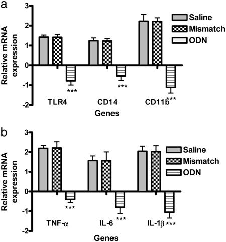
Neuropathic pain remains a prevalent and persistent clinical problem because of our incomplete understanding of its pathogenesis. This study demonstrates for the first time, to our knowledge, a critical role for CNS innate immunity by means of microglial Toll-like receptor 4 (TLR4) in the induction phase of behavioral hypersensitivity in a mouse and rat model of neuropathy. We hypothesized that after L5 nerve transection, CNS neuroimmune activation and subsequent cytokine expression are triggered by the stimulation of microglial membrane-bound TLR4. To test this hypothesis, experiments were undertaken to assess tactile and thermal hypersensitivity in genetically altered (i.e., TLR4 knockout and point-mutant) mice after L5 nerve transection. In a complementary study, TLR4 antisense oligodeoxynucleotide (ODN) was administered intrathecally to L5 spinal nerve injured rats to reduce the expression of spinal TLR4. Both the genetically altered mice and the rats treated with TLR4 antisense ODN displayed significantly attenuated behavioral hypersensitivity and decreased expression of spinal microglial markers and proinflammatory cytokines as compared with their respective control groups. This finding shows that TLR4 contributes to the initiation of CNS neuroimmune activation after L5 nerve transection. Further understanding of this early, specific, innate CNS/microglial response and how it leads to sustained glial/neuronal hypersensitivity may point to new therapies for the prevention and treatment of neuropathic pain syndromes.


Figures

Fig. 1.
Fig. 1.
Mechanical allodynia and thermal hyperalgesia in the L5 spinal nerve-transected mice. Shown are responses measured by foot-lift response frequency to stimulation with 0.008-g (a) and 0.015-g (b) von Frey filament. Paw-withdrawal latency (c) and tail-flick latency (d) are shown postoperatively. Mechanical allodynia and thermal hyperalgesia were significantly attenuated in KO mice (C57BL/10SCNJ; n = 21) and TLR4 mutant mice (C3H/HeJ; n = 8) relative to their respective controls, i.e., TLR4 wild-type mice (C57BL10/ScSnJ; n = 15) and C3H/HeN mice (n = 7). In a and b, asterisks indicate significant attenuation in tactile allodynia compared with the wild-type controls (***, P < 0.001; two-way ANOVA followed by Bonferroni post hoc test). In c and d, asterisks and ξ indicate significant decreases in paw-withdrawal and tail-flick latencies compared with wild type (***, P < 0.001; **, P < 0.01; *, P < 0.05; ξ, P < 0.05 vs. ScSnJ, two-way ANOVA followed by Bonferroni post hoc test). For a and b, results are reported as the mean response frequency from three trials of 10 stimulations each ± SEM. For c and d, the results are reported as the mean latency response time of three stimulations ± SEM.
Fig. 2.
Fig. 2.
Real-time quantitative RT-PCR analyses of mRNA temporal expression of microglial and astrocytic activation markers in mouse lumbar spinal cord. TaqMan real-time RT-PCR was performed. Total mRNA was isolated from L5 lumbar spinal cord tissue of KO mice (C57BL/10SCNJ) and TLR4 wild-type control mice (C57BL10/ScSnJ). Data are shown for normal, uninjured mice (N; n = 3 per strain) or KO mice (n = 4) and wild-type mice (n = 4) subjected to L5 nerve transection surgery at days 3, 7, and 14 for CD11b (a), CD14 (b), TLR4 (c) and GFAP (d) in the presence of their respective primers and probes. No PCR amplification was observed in the TLR4-deficient mice with TLR4 primers and probe (data not shown). (c) The time-course expression of TLR4 in wild-type mice after injury. The real-time PCR was performed in duplicate for both target gene and GAPDH. The level of gene expression was calculated after normalizing against GAPDH in each sample and is presented as relative mRNA expression units. Values are mean ± SEM. (**, P < 0.01 vs. KO; *, P < 0.05 vs. KO; two-way ANOVA followed by Bonferroni post hoc test.)
Fig. 3.
Fig. 3.
Real-time RT-PCR analyses of the temporal expression of proinflammatory cytokine mRNA in mouse lumbar spinal cord. Total mRNA was isolated from L5 lumbar spinal cord tissue of normal, uninjured mice (N) (n = 3 per strain), TLR4 KO mice (n = 4), and wild-type mice (n = 4) subjected to L5 nerve transection surgery at days 3, 7, and 14 for INF-γ (a), IL-1β (b) and TNF-α (c). The level of gene expression was calculated after normalizing against GAPDH in each sample and is presented as relative mRNA expression units. Values are mean ± SEM. (***, P < 0.001 vs. KO; *, P < 0.05 vs. KO; two-way ANOVA followed by Bonferroni post hoc test.)
Fig. 4.
Fig. 4.
Immunohistochemical staining for CD11b/CR3 of microglia in the ipsilateral L5 lumbar spinal cord dorsal horns 3, 7, and 14 days after surgery. Shown are representative photomicrographs depict microglial activation by using CD11b/CR3 immunoreactivity in uninjured TLR4 KO and its wild-type control mice and in L5 nerve transected KO and WT mice at 3, 7, and 14 days after surgery. Microglial activation in the dorsal horn of the spinal cord demonstrated a baseline staining in uninjured mice, a mild response at 3, 7, and 14 days after surgery for KO mice, and a moderate to intense response at day 3 and an intense response at 7 and 14 days after surgery in WT mice.
Fig. 5.
Fig. 5.
Dose–response relationship after daily intrathecal administration of TLR4 antisense ODN in L5 spinal nerve-transected rats. The effect of a daily intrathecal injection of two doses of TLR4 antisense ODN, mismatch ODN, or saline solution (10 or 20 μg per day) on mechanical allodynia (a) and paw-withdrawal latencies (b) is shown. Thermal and tactile hypersensitivity were significantly attenuated in a dose-dependent manner compared with saline and mismatch ODN-treated rats with marked attenuation observed with 10-μg daily injections of TLR4 antisense ODN starting at days 3–7 (P < 0.05 and P < 0.01) and day 1 (*, P < 0.05), day 3 (**, P < 0.01), and through day 7 (***, P < 0.001) for 20-μg daily injection of TLR4 antisense ODN (two-way ANOVA followed by Bonferroni post hoc test).
Fig. 6.
Fig. 6.
Effect of TLR4 antisense ODN on microglial activation and proinflammatory cytokine mRNA expression. A daily dose of 20 μg of TLR4 antisense ODN, mismatch ODN, or saline was injected intrathecally for 8 days, starting 1 day before L5 spinal nerve transection. The rats were killed on day 7 after surgery, and the lumbar spinal cord was processed for RT-PCR. RT-PCR revealed a significant down-regulation of TLR4 (P < 0.001). The down-regulation of TLR4 paralleled a significant decrease in the expression of two microglial activation markers, CD14 (***, P < 0.001) and CD11b (*** P < 0.001) (a), and in that of proinflammatory cytokines TNF-α, IL-6, and IL-1β (***, P < 0.001) (b). The level of gene expression was presented as a fold decrease relative to saline treated rats. Values were mean ± SEM. (n = 8 per treatment group; ***, P < 0.001; two-way ANOVA followed by Bonferroni post hoc test.)

References

    1. DeLeo, J. A. & Yezierski, R. P. (2001) Pain 90, 1–6. - PubMed
    1. DeLeo, J. A., Tanga, F. Y. & Tawfik, V. L. (2004) Neuroscientist 10, 40–52. - PubMed
    1. Vuong, C., Voyich, J. M., Fischer, E. R., Braughton, K. R., Whitney, A. R., DeLeo, F. R. & Otto, M. (2004) Cell. Microbiol. 6, 269–275. - PubMed
    1. Raghavendra, V., Tanga, F. Y. & DeLeo, J. A. (2004) Neuropsychopharmacology 29, 327–334. - PubMed
    1. Sommer, C., Galbraith, J. A., Heckman, H. M. & Myers, R. R. (1993) J. Neuropathol. Exp. Neurol. 52, 223–233. - PubMed

Publication types

MeSH terms