. 2005 Apr 12;102(15):5489-94.
doi: 10.1073/pnas.0402581102. Epub 2005 Apr 4.

Olfactory expression of a single and highly variable V1r pheromone receptor-like gene in fish species

Affiliations

Olfactory expression of a single and highly variable V1r pheromone receptor-like gene in fish species

Patrick Pfister et al. Proc Natl Acad Sci U S A. .

Abstract

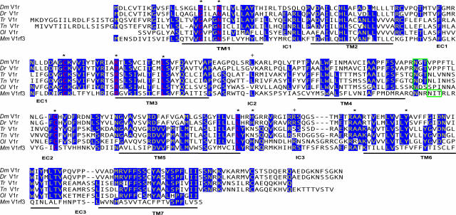
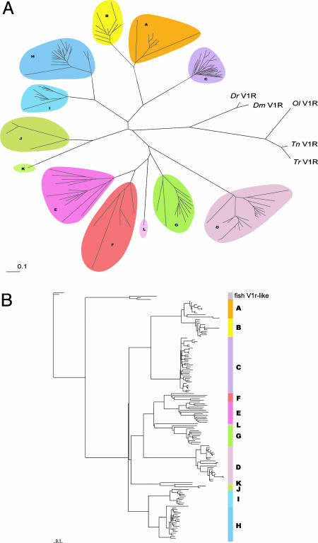
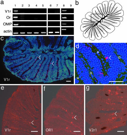
Sensory neurons expressing members of the seven-transmembrane V1r receptor superfamily allow mice to perceive pheromones. These receptors, which exhibit no sequence homology to any known protein except a weak similarity to taste receptors, have only been found in mammals. In the mouse, the V1r repertoire contains >150 members, which are expressed by neurons of the vomeronasal organ, a structure present exclusively in some tetrapod species. Here, we report the existence of a single V1r gene in multiple species of a non-terrestrial, vomeronasal organ-lacking taxon, the teleosts. In zebrafish, this V1r gene is expressed in chemosensory neurons of the olfactory rosette with a punctate distribution, strongly suggesting a role in chemodetection. This unique receptor gene exhibits a remarkably high degree of sequence variability between fish species. It likely corresponds to the original V1r present in the common ancestor of vertebrates, which led to the large and very diverse expansion of vertebrate pheromone receptor repertoires, and suggests the presence of V1rs in multiple nonmammalian phyla.


Figures

Fig. 1.
Fig. 1.
Fish V1r receptors. Alignment of the deduced amino acid of the zebrafish (Dr), giant danio (Dm), medaka (Ol), tetraodon (Tn), and fugu (Tr) V1r-like genes and the mouse (Mm) V1rf3 gene is shown. Conserved residues (at least four of six) are highlighted in blue. The 10 residues that are found in virtually all mouse V1rs are indicated by asterisks. The corresponding conserved residues in fish sequences are red. The green box indicates the position of the conserved N-linked glycosylation sites (NXS/T). +, the positions corresponding to the polymorphic variants found in the Tü, AB, and WIK Dr strains.
Fig. 2.
Fig. 2.
The fish and mouse V1r repertoires. (A) An unrooted tree representing V1r-like sequences from the five fish species together with the complete mouse V1r repertoire (the 12 families). Amino acid sequences were used for the generation of this tree. A comparable tree was generated by using nucleotide sequences (see Fig. 6). (B) Rooted tree, including all mouse V1r nucleotide sequences and the five fish sequences (Dr was used as an out-group). The corresponding tree for proteins is shown in Fig. 5.
Fig. 3.
Fig. 3.
Expression of V1r-like transcripts in the zebrafish olfactory system. (a) Expression of Dr V1r mRNA. cDNA from different adult tissues was amplified by PCR with specific primers for DrV1r, odorant receptor 1 (OR), olfactory marker protein (OMP), and actin. Lane 1, olfactory rosette; lane 2, gills; lane 3, olfactory bulb; lane 4, brain; lane 5, heart; lane 6, barbels; lane 7, lips; lane 8, genomic DNA; lane 9, olfactory rosette minus reverse transcriptase. Because no introns are found in the coding sequence of V1r and OMP, the sizes of the cDNA amplicons correspond to the size of the genomic amplicons. The amplicons corresponding to actin amplified from genomic material contain an intron and are therefore larger than cDNA amplicons. Exclusive V1r expression is observed in the olfactory rosette. (b) Drawing of a horizontal section of an olfactory rosette (lamellae are cut perpendicular to their flat face). The gray zone in the lamellae, close to the center, indicates the location of the sensory neuroepithelium. The black lines correspond to the cartilage. The rectangle indicates the area shown in d. (c and d) In situ hybridizations of horizontal sections through the olfactory rosette, with an antisense DrV1r-like RNA probe. Neurons expressing V1r-like mRNA appear as fluorescent red. The blue color corresponds to DAPI staining of the nuclei. The asterisks and squares indicate the lumen between the lamellae and the extracellular matrix, respectively. (e–g) In situ hybridizations with V1r, OR1, and V2r1 probes, respectively. The numerous neurons positive for V2r1 likely reflect crosshybridization with multiple V2r sequences. (Scale bar, 30 μm.) Arrowheads indicate some of the labeled neurons.

Similar articles

Cited by

References

    1. Buck, L. & Axel, R. (1991) Cell 65, 175–187. - PubMed
    1. Ryba, N. J. & Tirindelli, R. (1997) Neuron 19, 371–379. - PubMed
    1. Matsunami, H. & Buck, L. B. (1997) Cell 90, 775–784. - PubMed
    1. Herrada, G. & Dulac, C. (1997) Cell 90, 763–773. - PubMed
    1. Speca, D. J., Lin, D. M., Sorensen, P. W., Isacoff, E. Y., Ngai, J. & Dittman, A. H. (1999) Neuron 23, 487–498. - PubMed

Publication types

LinkOut - more resources