

Chromic-P32 phosphate treatment of implanted pancreatic carcinoma: mechanism involved
- PMID: 15810075
- PMCID: PMC4305778
- DOI: 10.3748/wjg.v11.i14.2101
Chromic-P32 phosphate treatment of implanted pancreatic carcinoma: mechanism involved
Abstract
Aim: To study the effects of chromic-P32 phosphate (32P colloids) interstitial administration in Pc-3 implanted pancreatic carcinoma, and investigate its anticancer mechanism.
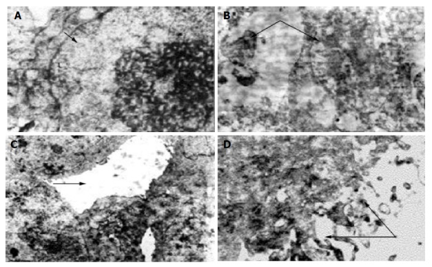
Methods: Ninety-eight tumor bearing nude mice were killed at different time points after the injection of 32P colloids to the tumor core with observed radioactivity. The light microscopy, transmission electron microscopy (TEM) and immuno-histochemistry and flow cytometry were used to study the rates of tumor cell necrosis, proliferating cell nuclear antigen index, the micro vessel density (MVD). The changes of the biological response to the lymphatic transported 32P colloids in the inguinal lymph node (ILN) were dynamically observed, and the percentage of tumor cell apoptosis, and Apo2.7, caspase-3, Bcl-2, Bax-related gene expression were observed too.
Results: The half-life of effective medication is 13 d after injection of 32P colloids to the tumor stroma, in 1-6 groups, the tumor cell necrosis rates were 20%, 45%, 65%, 70%, 95% and 4%, respectively (F = 4.14-105.36, P<0.01). MVD were 38.5+/-4.0, 28.0+/-2.9, 17.0+/-2.9, 8.8+/-1.5, 5.7+/-2.3 and 65.0+/-5.2 (t = 11.9-26.1, P<0.01), respectively. Under TEM fairly differentiated Pc-3 cells were found. Thirty days after medication, tumors were shrunk and dried with scabs detached, and those in control group increased in size prominently with plenty of hypodermic blood vessels. In all animals the ILN were enlarged but in medicated animals they appeared later and smaller than those in control group. The extent of irradiative injury in ILN was positively correlated to the dosage of medication. Typical tumor cell apoptosis could be found under TEM in animals with intra-tumoral injection of low dosed 32P colloids. The peak of apoptosis occurred in 2.96 MBq group and 24 h after irradiation. In the course of irradiation-induced apoptosis, the value of Bcl-2/Bax was down regulated; Apo2.7 and caspase-3 protein expression were prominently increased dose dependently.
Conclusion: 32P colloids intra-tumor injection having prominent anticancer effectiveness may reveal the ability of promoting cell differentiation. The low dose 32P colloids may induce human pancreatic carcinoma Pc-3 implanted tumor cell apoptosis; Apo2.7, caspase-3, Bcl-2 and Bax protein participated in regulating the process of irradiation induced cell apoptosis.
Figures


Similar articles
-
Intratumoral injection of 32P-chromic phosphate in the treatment of implanted pancreatic carcinoma.Cancer Biother Radiopharm. 2010 Apr;25(2):215-24. doi: 10.1089/cbr.2008.0596. Cancer Biother Radiopharm. 2010. PMID: 20423235
-
Interstitial chemotherapy with ricin-loaded thermosensitive hydrogel in pancreatic cancer xenograft.Hepatobiliary Pancreat Dis Int. 2009 Aug;8(4):418-23. Hepatobiliary Pancreat Dis Int. 2009. PMID: 19666413
-
Anti-tumor activities and apoptosis-regulated mechanisms of bufalin on the orthotopic transplantation tumor model of human hepatocellular carcinoma in nude mice.World J Gastroenterol. 2007 Jun 28;13(24):3374-9. doi: 10.3748/wjg.v13.i24.3374. World J Gastroenterol. 2007. PMID: 17659679 Free PMC article.
-
³²P-chromic phosphate-Poly(L-Lactide) seeds of sustained release and their brachytherapy for prostate cancer with lymphatic metastasis.Cancer Biother Radiopharm. 2012 Sep;27(7):446-51. doi: 10.1089/cbr.2011.1141. Epub 2012 Jul 26. Cancer Biother Radiopharm. 2012. PMID: 22834706
-
[Mechanisms of inhibition of growth of human pancreatic carcinoma implanted in nude mice by somatostatin receptor subtype 2].Zhonghua Yi Xue Za Zhi. 2004 May 2;84(9):760-5. Zhonghua Yi Xue Za Zhi. 2004. PMID: 15200918 Chinese.
Cited by
-
[Experimental study of CT guided ³²P-CP-PLLA microparticle implantation in the treatment of rabbit VX2 lung tumor].Zhongguo Fei Ai Za Zhi. 2011 Jan;14(1):1-6. doi: 10.3779/j.issn.1009-3419.2011.01.01. Zhongguo Fei Ai Za Zhi. 2011. PMID: 21219823 Free PMC article. Chinese.
-
Short-term effectiveness of radiochemoembolization for selected hepatic metastases with a combination protocol.World J Gastroenterol. 2012 Oct 7;18(37):5249-59. doi: 10.3748/wjg.v18.i37.5249. World J Gastroenterol. 2012. PMID: 23066320 Free PMC article.
References
-
- Tang ZY. Modern Oncology. 2nd Ed. Shanghai: Shanghai University of Medical Science Press; 2000. pp. 1132–1134.
-
- Qin MX, Diao GP. Tianjin Scientific Technology Press 1996: 11-13. Clinical Interventional Nuclear Medicine. 1st Ed.
-
- Shen K, Nirmal L, Han Q, Wu J, Lu J, Zhang J, Liu G, Shao Z, Shen Z. Sentinel lymph node biopsy in breast cancer. Zhonghua WaiKe ZaZhi. 2002;40:347–350. - PubMed
-
- Pattillo RA, Collier BD, Abdel-Dayem H, Ozker K, Wilson C, Ruckert AC, Hamilton K. Phosphorus-32-chromic phosphate for ovarian cancer: I. Fractionated low-dose intraperitoneal treatments in conjunction with platinum analog chemotherapy. J Nucl Med. 1995;36:29–36. - PubMed
Publication types
MeSH terms
Substances
LinkOut - more resources
Full Text Sources
Medical
Research Materials
