. 2005 Apr;71(4):2095-105.
doi: 10.1128/AEM.71.4.2095-2105.2005.

Analysis of fluorescent protein expression in transformants of Rickettsia monacensis, an obligate intracellular tick symbiont

Affiliations

Analysis of fluorescent protein expression in transformants of Rickettsia monacensis, an obligate intracellular tick symbiont

Gerald D Baldridge et al. Appl Environ Microbiol. 2005 Apr.

Abstract

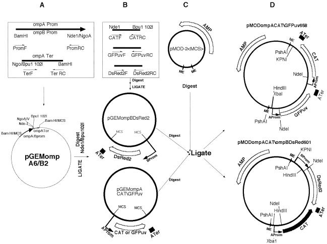
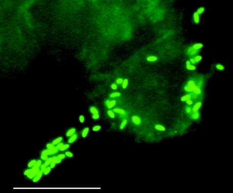
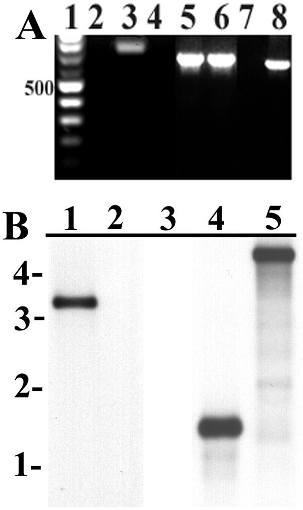
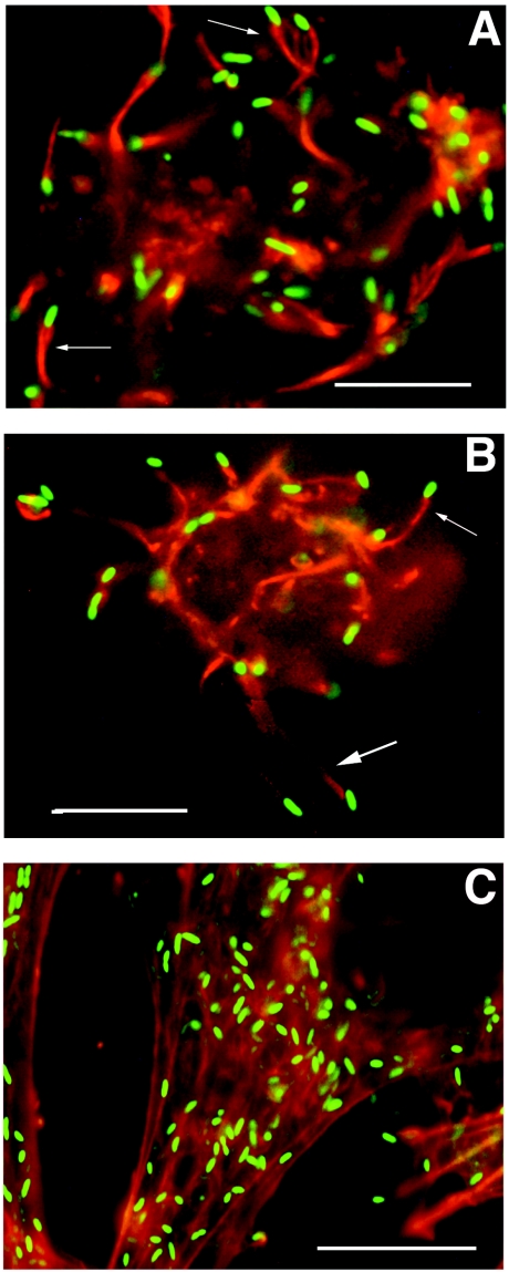
We developed and applied transposon-based transformation vectors for molecular manipulation and analysis of spotted fever group rickettsiae, which are obligate intracellular bacteria that infect ticks and, in some cases, mammals. Using the Epicentre EZ::TN transposon system, we designed transposons for simultaneous expression of a reporter gene and a chloramphenicol acetyltransferase (CAT) resistance marker. Transposomes (transposon-transposase complexes) were electroporated into Rickettsia monacensis, a rickettsial symbiont isolated from the tick Ixodes ricinus. Each transposon contained an expression cassette consisting of the rickettsial ompA promoter and a green fluorescent protein (GFP) reporter gene (GFPuv) or the ompB promoter and a red fluorescent protein reporter gene (DsRed2), followed by the ompA transcription terminator and a second ompA promoter CAT gene cassette. Selection with chloramphenicol gave rise to rickettsial populations with chromosomally integrated single-copy transposons as determined by PCR, Southern blotting, and sequence analysis. Reverse transcription-PCR and Northern blots demonstrated transcription of all three genes. GFPuv transformant rickettsiae exhibited strong fluorescence in individual cells, but DsRed2 transformants did not. Western blots confirmed expression of GFPuv in R. monacensis and in Escherichia coli, but DsRed2 was expressed only in E. coli. The DsRed2 gene, but not the GFPuv gene, contains many GC-rich amino acid codons that are rare in the preferred codon suite of rickettsiae, possibly explaining the failure to express DsRed2 protein in R. monacensis. We demonstrated that our vectors provide a means to study rickettsia-host cell interactions by visualizing GFPuv-fluorescent R. monacensis associated with actin tails in tick host cells.


Figures

FIG. 1.
FIG. 1.
Construction of rickettsial transposon vectors. (A) PCR amplification of R. rickettsii Hlp#2 ompA and ompB promoters with forward (PromF) and reverse complement (PromRC) primers and of the ompA transcription terminator with TerF and TerRC primers, all containing end-terminal restriction endonuclease sites (Table 1). Amplicons were digested with BamHI and NgoAIV and ligated into the pGem3Z BamHI site within the MCS to obtain pGEMompA6 and -ompB2. (B) PCR amplification of CAT, GFPuv, and DsRed2 gene open reading frames with forward (F) NdeI and reverse complement (RC) BpuI 102I end-terminal primers. Amplicons and pGEMompA6 and -B2 plasmids were digested with the same enzyme pair and ligated to produce pGEMompACAT or -GFPuv and pGEMompBDsRed2. (C) The Epicentre pMOD-2-<MCS> transposon vector was cleaved at restriction sites within the MCS and ligated with two rickettsial gene expression cassettes (ompACAT and ompAGPPuv or ompBDsRed2) removed (see text) from the plasmids shown in panel B. (D) Final vectors, pMODompACAT\GFPuv658 (pMOD658) and pMODompACAT\ompBDsRed601 (pMOD601), showing relative positions of 19-nucleotide mosaic element (ME) sites recognized by EZ::TN transposase, ompA and -B promoters (Aprom and Bprom), the transcription terminators (ATer), and the CAT, Discoma red fluorescent protein (DsRed2), green fluorescent protein (GFPuv), and ampicillin resistance (AMP) genes, with directions of transcription indicated by arrows. Lines indicate positions of restriction enzyme sites involved in Southern blot experiments, plasmid rescue cloning, and excision of the transposons (PshA1) from the vectors for incubation with EZ::TN transposase to form transposome complexes for electroporation.
FIG. 2.
FIG. 2.
GFPuv-fluorescent R. monacensis transformant cells (Rmona658) in tick ISE6 cells. Bar, 10 μm. Magnification, ×1,000, with a fluorescein isothiocyanate filter.
FIG. 3.
FIG. 3.
Detection of transposons in Rmona658 and Rmona601 DNA extracts. (A) PCR amplicons electrophoresed on a 1% agarose gel stained with EtBr. Lane 1 contains a 100-bp marker ladder, with the 500-bp band indicated at left. GFP primer amplicons from untransformed control R. monacensis (lane 2) and Rmona658 (lane 3); CAT primer amplicons from controls (lane 4), Rmona658 (lane 5), and Rmona601 (lane 6); DsRed2 primer amplicons from controls (lane 7) and Rmona601 (lane 8). (B) Southern blots with gel migration positions of a 1-kb marker ladder indicated at left. HindIII-digested DNA from Rmona658 (lane 1) or untransformed controls (lane 2) hybridized with a GFPuv probe; NdeI-digested DNA from controls (lane 3) or Rmona601 (lane 4) and KpnI-digested DNA from Rmona601 (lane 5) hybridized with a DsRed 2 probe.
FIG. 4.
FIG. 4.
Detection of CAT, GFPuv, and DsRed2 mRNAs in Rmona658 and Rmona601 RNA extracts by RT-PCR and Northern blotting. (A) Rmona658 RT-PCR analysis. Lane 1: 100-bp marker ladder, with the 700-bp band indicated at left (asterisk). Rmona658 no-RT control reactions with CAT (lane 2) and GFPuv (lane 3) primers, respectively. Rmona658 (lane 4) and untransformed control (lane 5) RT-PCRs with CAT primers. Rmona658 (lane 6) and untransformed control (lane 7) RT-PCRs with GFPuv primers. (B) Rmona601 RT-PCR analysis. Lane 1: 100-bp marker as described above. Untransformed control (lane 2) and Rmona601 (lane 3) RT-PCRs with CAT primers. Untransformed control (lane 4) and Rmona601 (lane 5) RT-PCRs with DsRed2 primers. Rmona601 no-RT control reactions with CAT (lane 6) and DsRed2 (lane 7) primers. (C) Northern blot. Lane 1: 0.5- and 1.0-kb RNA markers. Rmona658 (lane 2) and untransformed control (lane 3) RNA extracts hybridized with GFPuv probe. Rmona658 (lane 4), untransformed control (lane 5), and Rmona601 (lane 6) RNA extracts hybridized with CAT probe. Untransformed control (lane 7) and Rmona601 (lane 8) RNA extracts hybridized with DsRed2 probe.
FIG. 5.
FIG. 5.
Western immunoblots of R. monancensis, E. coli, and RF/6A cell protein extracts. (A) GFP immunoblot. Lane 1: SeeBlue Plus 2 markers, with sizes in kilodaltons indicated at left. Lane 2: Rmona658 extract. Lane 3: pMOD658 transformant E. coli extract. Lane 4: untransformed R. monacensis extract. Lane 5: untransformed E. coli extract. (B) DsRed immunoblot. Lane 1: SeeBlue Plus 2 markers. Lane 2: Rmona601 extract. Lane 3: untransformed R. monacensis extract. Lane 4: DsRed2 transformant RF/6A extract. Lane 5: untransformed E. coli extract. Lane 6: pMOD601 transformant E. coli extract.
FIG. 6.
FIG. 6.
Visualization of GFPuv-fluorescent rickettsiae associated with rhodamine-labeled (red) host cell actin structures. (A and B) Rickettsiae (green rods) in tick IDE8 cells associated with actin tail structures (arrows) that were primarily cytosolic (A) or in pseudopodia (B). (C) Rickettsiae in vertebrate RF6A cells. Bar, 10 μm.

Similar articles

Cited by

References

    1. Anderson, B. E., G. A. McDonald, D. C. Jones, and R. L. Regnery. 1990. A protective protein antigen of Rickettsia rickettsii has tandemly repeated, near-identical sequences. Infect. Immun. 58:2760-2769. - PMC - PubMed
    1. Andersson, S. G. E., and P. M. Sharp. 1996. Codon usage and base composition in Rickettsia prowazekii. J. Mol. Evol. 42:525-536. - PubMed
    1. Andersson, S. G. E., A. Zomorodipour, J. O. Andersson, T. Sicheritz-Ponten, U. C. M. Alsmark, R. M. Podowski, A. K. Naslund, A. Eriksson, H. H. Winkler, and C. G. Kurland. 1998. The genome sequence of Rickettsia prowazekii and the origin of mitochondria. Nature 396:133-140. - PubMed
    1. Anonymous. 2001. Living colors DsRed2. Clonetechniques 16:2-3.
    1. Azad, A. F., and C. B. Beard. 1998. Rickettsial pathogens and their arthropod vectors. Emerg. Infect. Dis. 4:179-186. - PMC - PubMed

Publication types

Substances

LinkOut - more resources