. 2005 Apr 25;92(8):1450-8.
doi: 10.1038/sj.bjc.6602518.

Immunosuppressive effects of radiation on human dendritic cells: reduced IL-12 production on activation and impairment of naive T-cell priming

Affiliations

Immunosuppressive effects of radiation on human dendritic cells: reduced IL-12 production on activation and impairment of naive T-cell priming

A Merrick et al. Br J Cancer. .

Abstract

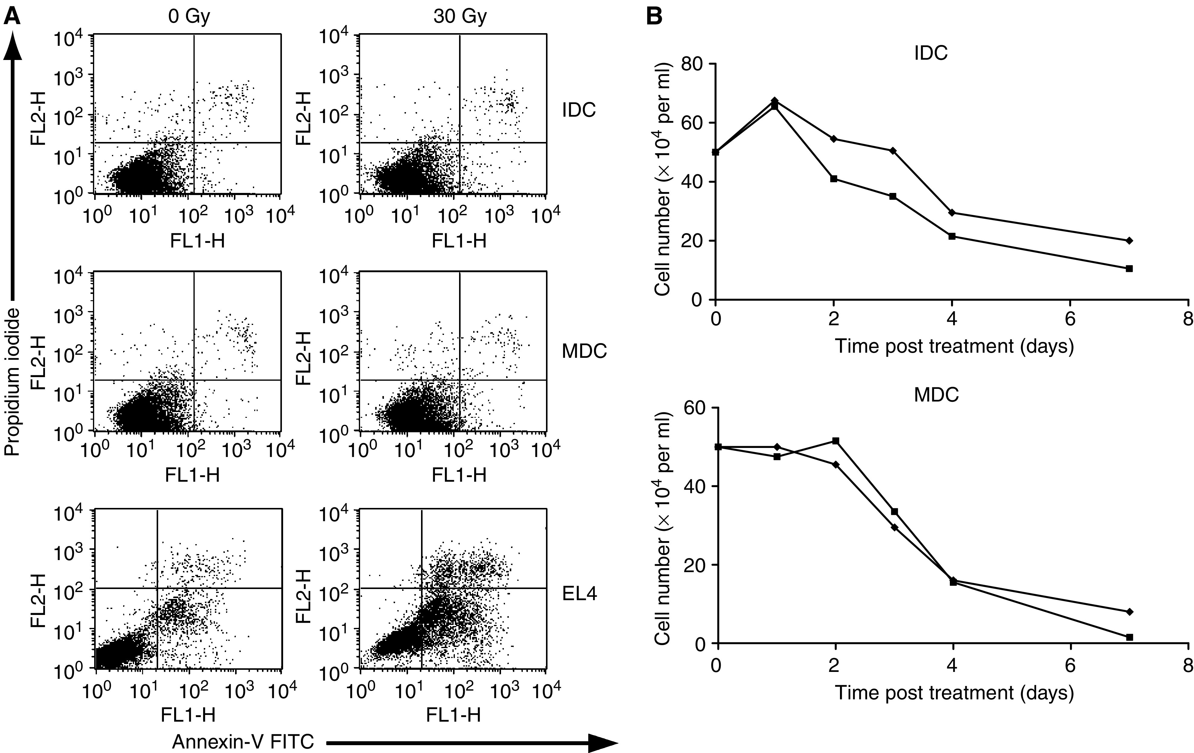
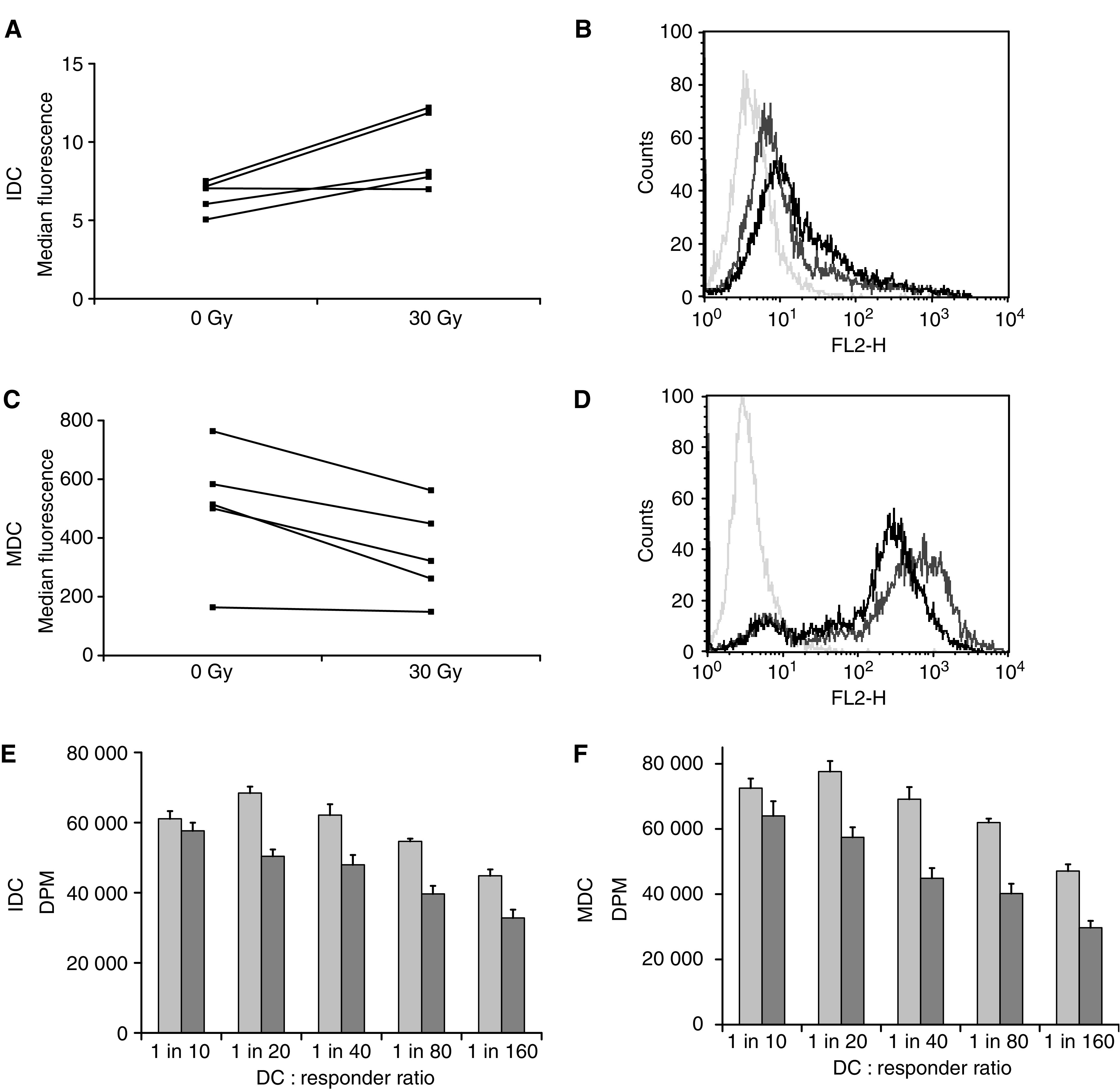
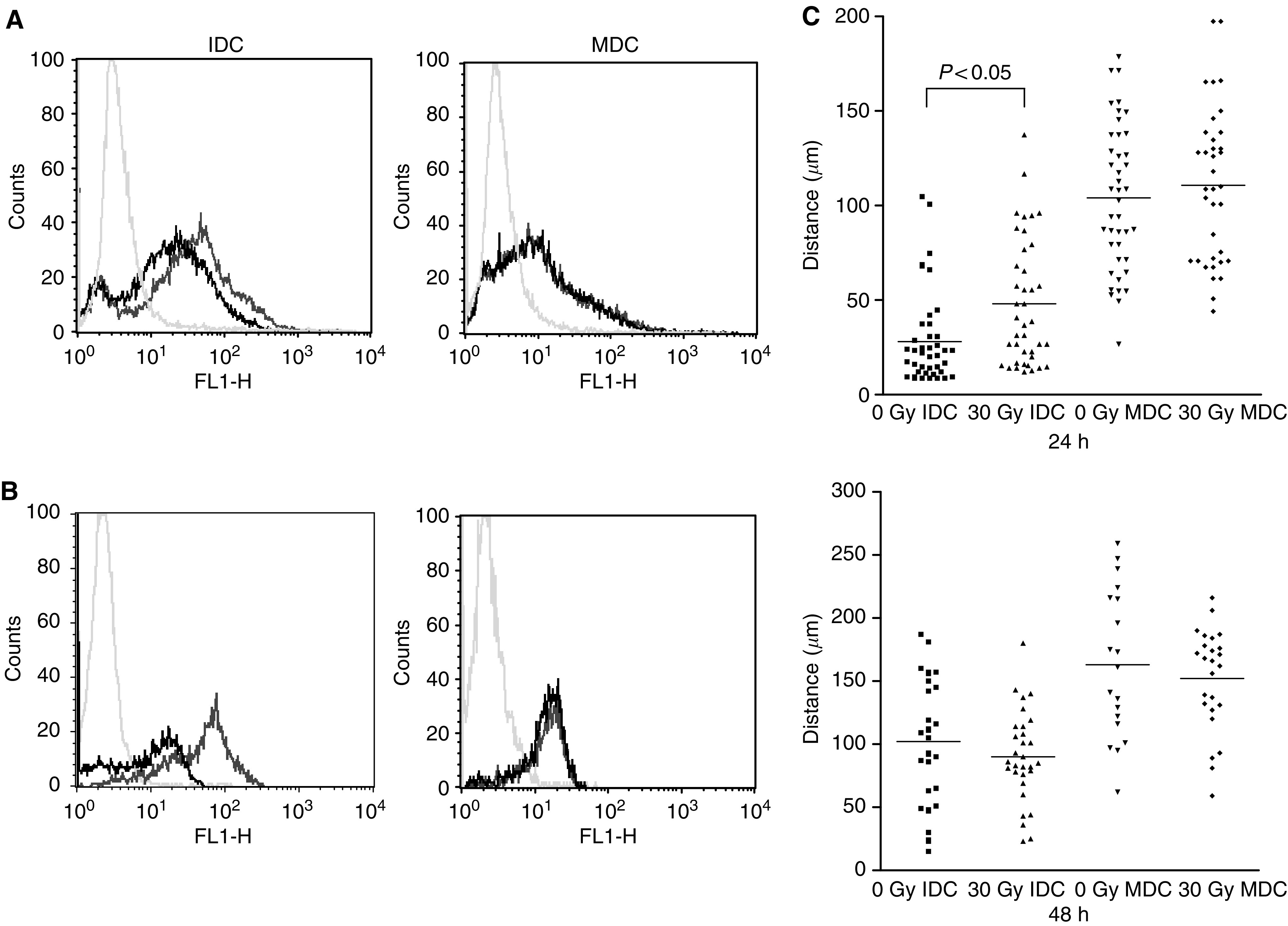
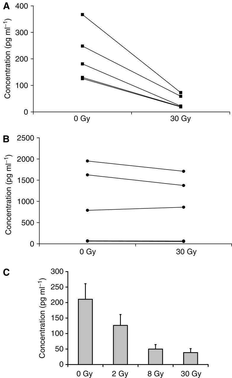
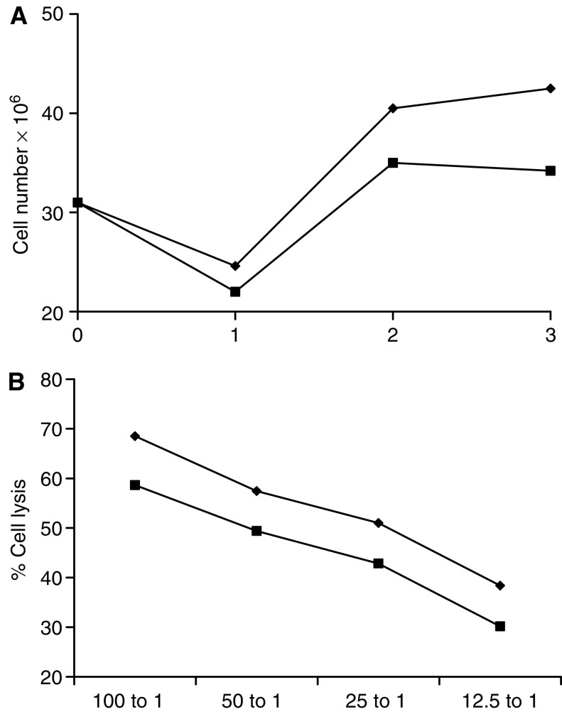
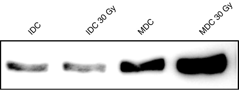
Dendritic cells (DC) are professional antigen-presenting cells (APC) of the immune system, uniquely able to prime naive T-cell responses. They are the focus of a range of novel strategies for the immunotherapy of cancer, a proportion of which include treating DC with ionising radiation to high dose. The effects of radiation on DC have not, however, been fully characterised. We therefore cultured human myeloid DC from CD14+ precursors, and studied the effects of ionising radiation on their phenotype and function. Dendritic cells were remarkably resistant against radiation-induced apoptosis, showed limited changes in surface phenotype, and mostly maintained their endocytic, phagocytic and migratory capacity. However, irradiated DC were less effective in a mixed lymphocyte reaction, and on maturation produced significantly less IL-12 than unirradiated controls, while IL-10 secretion was maintained. Furthermore, peptide-pulsed irradiated mature DC were less effective at naive T-cell priming, stimulating fewer effector cells with lower cytotoxicity against antigen-specific targets. Hence irradiation of DC in vitro, and potentially in vivo, has a significant impact on their function, and may shift the balance between T-cell activation and tolerization in DC-mediated immune responses.


Figures

Figure 1
Figure 1
Dendritic cell death following radiation. Apoptosis/necrosis of DC was assessed by FACS analysis after incubation with Annexin-V FITC and propidium iodide (A). No significant difference was seen in the apoptotic (lower right quadrant) or necrotic (upper right quadrant) populations of IDC or MDC±30 Gy at 24 h post treatment, in contrast to the significant early apoptotic death of radiosensitive EL4. (B) Dendritic cell viability was further assessed by seeding cells at constant density, and counting live cells daily using trypan blue exclusion. No statistical difference was found in the rate of cell death between irradiated (▪) or nonirradiated (⧫) controls for IDC or MDC. Data shown are representative of five similar experiments.
Figure 2
Figure 2
CD86 expression and MLR activity of irradiated dendritic cells. Changes in surface phenotype of DC were assessed 24 h post treatment by FACS analysis. Expression of the co-stimulatory molecule CD86 was significantly upregulated on IDC after irradiation (P<0.04), and downregulated on MDC (P<0.02), though levels on remained >10 fold less on IDC vs MDC. Data shown as MFI of five donors (A, C), and as a representative histogram for one donor (B, D). Mixed lymphocyte reaction: irradiated (dark bars) or control (light bars) IDC (E) and MDC (F) were incubated with responder allogeneic T cells at different ratios and incubated for 5 days before the addition of 3H-thymidine for 18 h, followed by harvesting and counting. Data are representative of four similar experiments.
Figure 3
Figure 3
Endocytosis, phagocytosis and motility of irradiated DC. Fluid-phase endocytosis by DC was measured following incubation with BODIPY-labelled BSA (A). Dendritic cell were incubated with the dye for 30 min at either 37°C or at 0°C as a negative control (pale grey histogram). Uptake shown by shift in fluorescence for unirradiated DC (dark grey histogram) vs irradiated DC (black histogram). Phagocytosis was similarly measured by uptake of fluorescently labelled whole tumour cells, with untreated DC alone as the control (B). These data are representative of three similar experiments. (C) Tracking (30-min) of IDC and MDC 24 and 48 h post irradiation. Each data point represents the distance travelled by an individual cell, with the horizontal bar showing the mean for the group.
Figure 4
Figure 4
Cytokine secretion by activated DC on irradiation. Cytokine secretion by LPS-activated DC was assayed by ELISA. IL-12 p70 secretion was significantly reduced on irradiation to 30 Gy of DC from five separate donors (P<0.001) (A), while IL-10 was unaffected (B). Pooled results show a dose response in IL-12 secretion at 2, 8, and 30 Gy (C).
Figure 5
Figure 5
Priming of a naïve cytotoxic T-cell response by irradiated DC. Irradiated (30 Gy) or control MDC were pulsed with MART-1 peptide and incubated with PBMC. T-cell cultures were re-stimulated twice at weekly intervals and assayed for CTL activity at 21 days. Weekly cell counts showed less proliferation of T cells incubated with irradiated (▪) than unirradiated (⧫) DC (A shows a representative donor). CTL lysis of peptide-pulsed targets was lower in cultures primed with irradiated DC (square), than untreated controls (diamond) (B). These data are shown as pooled results of CTL killing of MART-1 pulsed targets for four separate donors. Unirradiated vs irradiated groups were statistically significant (P<0.001).
Figure 6
Figure 6
Nuclear RelB expression by irradiated dendritic cells. Nuclear extracts were prepared from irradiated and control IDC and MDC, and RelB expression analysed by Western blot.

References

    1. Anton D, Dabadghao S, Palucka K, Holm G, Yi Q (1998) Generation of dendritic cells from peripheral blood adherent cells in medium with human serum. Scand J Immunol 47: 116–121 - PubMed
    1. Banchereau J, Steinman RM (1998) Dendritic cells and the control of immunity. Nature 392: 245–252 - PubMed
    1. Cao MD, Chen ZD, Xing Y (2004) Gamma irradiation of human dendritic cells influences proliferation and cytokine profile of T cells in autologous mixed lymphocyte reaction. Cell Biol Int 28: 223–228 - PubMed
    1. Celluzzi CM, Falo LD (1998) Physical interaction between dendritic cells and tumour cells results in an immunogen that induces protective and therapeutic tumour rejection. J Immunol 160: 3081–3085 - PubMed
    1. Chakravarty PK, Alfieri A, Thomas EK, Beri V, Tanaka KE, Vikram B, Guha C (1999) Flt3-ligand administration after radiation therapy prolongs survival in a murine model of metastatic lung cancer. Cancer Res 59: 6028–6032 - PubMed