

Nanopattern-induced changes in morphology and motility of smooth muscle cells
- PMID: 15814139
- PMCID: PMC2376810
- DOI: 10.1016/j.biomaterials.2005.01.058
Nanopattern-induced changes in morphology and motility of smooth muscle cells
Abstract
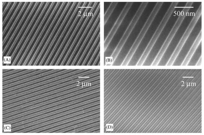
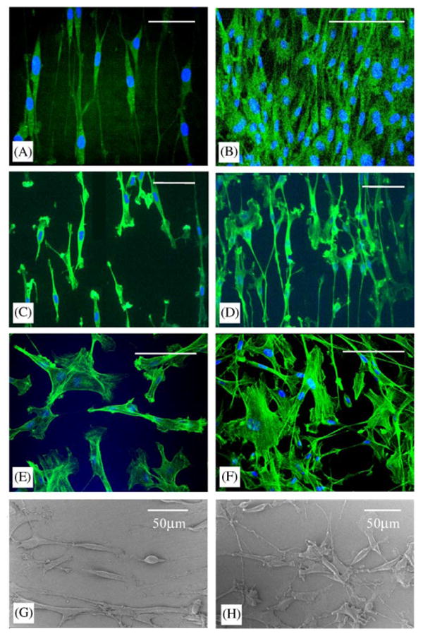
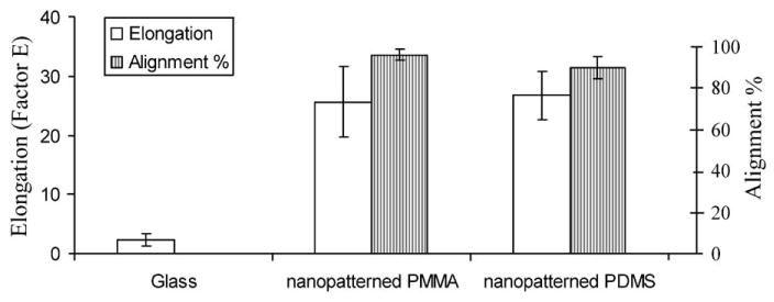
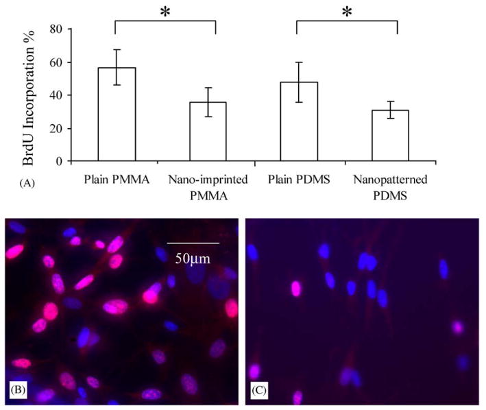
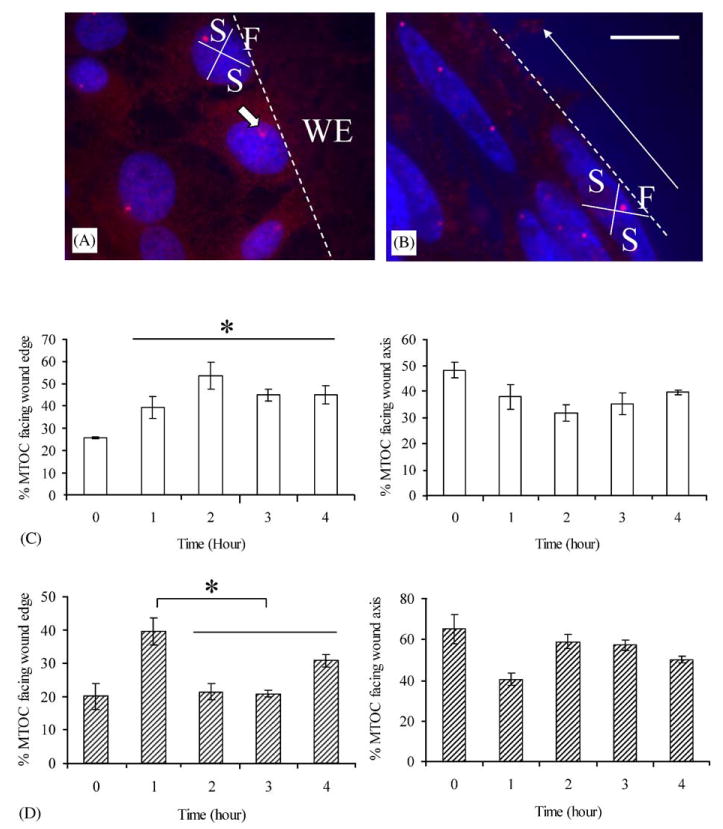
Cells are known to be surrounded by nanoscale topography in their natural extracellular environment. The cell behavior, including morphology, proliferation, and motility of bovine pulmonary artery smooth muscle cells (SMC) were studied on poly(methyl methacrylate) (PMMA) and poly(dimethylsiloxane) (PDMS) surfaces comprising nanopatterned gratings with 350 nm linewidth, 700 nm pitch, and 350 nm depth. More than 90% of the cells aligned to the gratings, and were significantly elongated compared to the SMC cultured on non-patterned surfaces. The nuclei were also elongated and aligned. Proliferation of the cells was significantly reduced on the nanopatterned surfaces. The polarization of microtubule organizing centers (MTOC), which are associated with cell migration, of SMC cultured on nanopatterned surfaces showed a preference towards the axis of cell alignment in an in vitro wound healing assay. In contrast, the MTOC of SMC on non-patterned surfaces preferentially polarized towards the wound edge. It is proposed that this nanoimprinting technology will provide a valuable platform for studies in cell-substrate interactions and for development of medical devices with nanoscale features.
Figures
References
-
- Nerem RM. Tissue engineering: confronting the transplantation crisis. Adv Exp Med Biol. 2003;534:1–9. - PubMed
-
- Langer R, Vacanti JP. Tissue engineering. Science. 1993;260(5110):920–6. - PubMed
-
- Patel N, Padera R, Sanders GH, Cannizzaro SM, Davies MC, Langer R, et al. Spatially controlled cell engineering on biodegradable polymer surfaces. Faseb J. 1998;12(14):1447–54. - PubMed
-
- Boyan BD, Hummert TW, Dean DD, Schwartz Z. Role of material surfaces in regulating bone and cartilage cell response. Biomaterials. 1996;17(2):137–46. - PubMed
-
- Aucoin L, Griffith CM, Pleizier G, Deslandes Y, Sheardown H. Interactions of corneal epithelial cells and surfaces modified with cell adhesion peptide combinations. J Biomater Sci Polym Ed. 2002;13(4):447–62. - PubMed
Publication types
MeSH terms
Substances
Grants and funding
LinkOut - more resources
Full Text Sources
Other Literature Sources
