Comparative Study
. 2005 Apr 6;25(14):3701-11.
doi: 10.1523/JNEUROSCI.4346-04.2005.

Matrix metalloproteinase-3: a novel signaling proteinase from apoptotic neuronal cells that activates microglia

Affiliations
Comparative Study

Matrix metalloproteinase-3: a novel signaling proteinase from apoptotic neuronal cells that activates microglia

Yoon Seong Kim et al. J Neurosci. .

Abstract

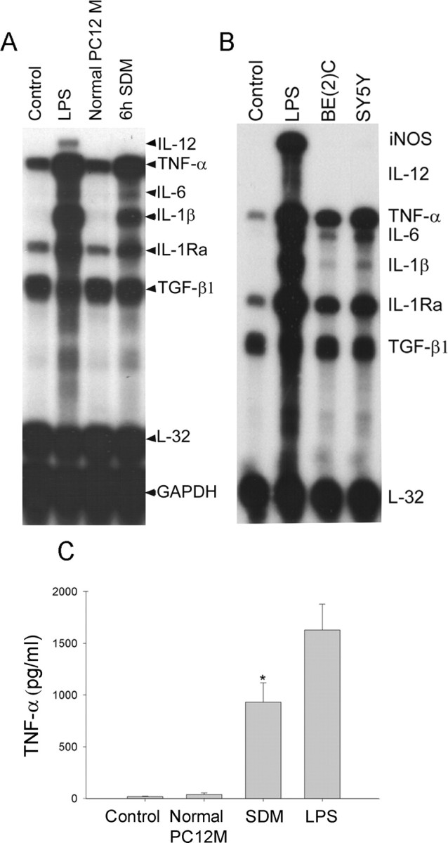
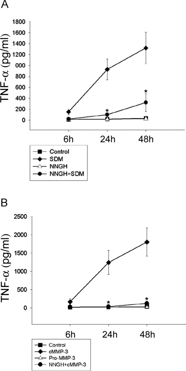
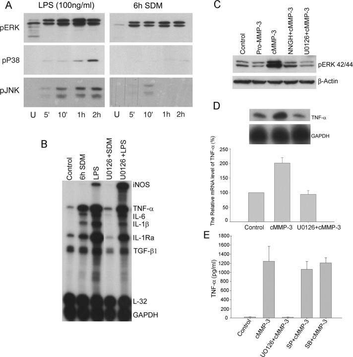
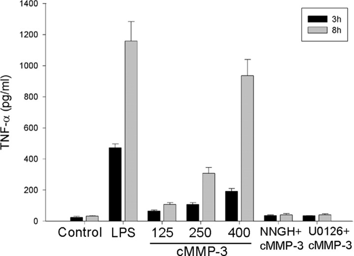
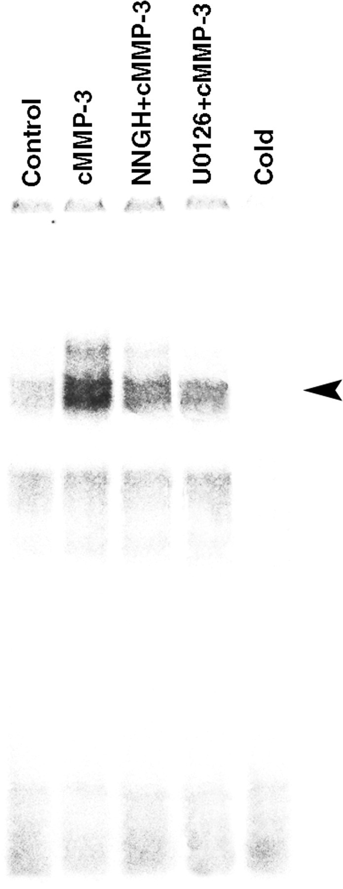
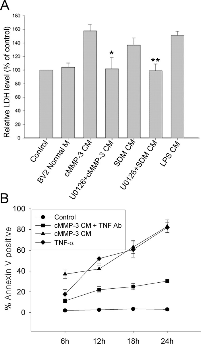
Microglial activation and inflammation are associated with progressive neuronal apoptosis in neurodegenerative human brain disorders. We sought to investigate molecular signaling mechanisms that govern activation of microglia in apoptotic neuronal degeneration. We report here that the active form of matrix metalloproteinase-3 (MMP-3) was released into the serum-deprived media (SDM) of PC12 cells and other media of apoptotic neuronal cells within 2-6 h of treatment of the cells, and SDM and catalytic domain of recombinant MMP-3 (cMMP-3) activated microglia in primary microglia cultures as well as BV2 cells, a mouse microglia cell line. Both SDM and cMMP-3 induced generation of tumor necrosis factor alpha (TNF-alpha), interleukin-6 (IL-6), IL-1beta, and interleukin-1 receptor antagonist but not IL-12 and inducible nitric oxide synthase, which are readily induced by lipopolysaccharide, in microglia, suggesting that there is a characteristic pattern of microglial cytokine induction by apoptotic neurons. Neither glial cell line-derived neurotrophic factor nor anti-inflammatory cytokines, such as IL-10 and transforming growth factor-beta1, were induced. SDM and cMMP-3 extensively released TNF-alpha from microglia and activated the nuclear factor-kappaB pathway, and these microglial responses were totally abolished by preincubation with an MMP-3 inhibitor, NNGH [N-isobutyl-N-(4-methoxyphenylsulfonyl)-glycylhydroxamic acid]. MMP-3-mediated microglial activation mostly depended on ERK (extracellular signal-regulated kinase) phosphorylation but not much on either JNK (c-Jun N-terminal protein kinase) or p38 activation. Conditioned medium of SDM- or cMMP-3-activated BV2 cells caused apoptosis of PC12 cells. These results strongly suggest that the distinctive signal of neuronal apoptosis is the release of active form of MMP-3 that activates microglia and subsequently exacerbates neuronal degeneration. Therefore, the release of MMP-3 from apoptotic neurons may play a major role in degenerative human brain disorders, such as Parkinson's disease.


Figures

Figure 1.
Figure 1.
The supernatants from apoptotic neuronal-mediated induction of cytokine genes in BV2 cells and TNF-α release. A, SDM from apoptotic PC12 culture strongly induced microglial cytokine genes. BV2 cells were treated for 6 h with either concentrated 6 h SDM (50 μg of proteins) or equivalents from normal PC12 culture. Various cytokine mRNA levels, including IL-12, TNF-α, IL-6, IL-1β, IL-1Ra, TGF-β1, and iNOS, were determined using RPA. L32 and GAPDH were used as internal controls. B, Supernatants from staurosporine-induced apoptotic SK-N-BE(2)C and SH-SY5Y also resulted in a similar pattern of cytokine induction in microglia. Apoptosis of both SK-N-BE(2)C and SH-SY5Y cells was induced by 4 h staurosporine treatment, and then, to remove staurosporine completely, cells were washed three times with fresh media. After the last wash, cells were incubated in fresh media for 6 h, and then supernatant was collected for microglial treatment. After 6 h treatment with supernatants, cytokine mRNA levels of BV2 cells were analyzed. Pictures shown are representative autoradiographs of the four independent RPAs. C, SDM caused TNF-α release from microglia. TNF-α levels were measured in the microglial culture media after 24 h treatments with each condition using ELISA. Values represent the mean ± SD (n = 4). Control, Untreated; Normal PC12 M, supernatant from PC12 cells in serum-containing normal medium for 6 h; SDM, supernatant from PC12 cells in serum-deprived medium for 6 h; LPS, 100 ng/ml LPS; SY5Y, supernatant from apoptotic SH-SY5Y cells that were treated with staurosporine (10 μm) for 4 h and then incubated with fresh medium for 6 h; BE(2)C, supernatant from apoptotic SK-N-BE(2)C cells that were treated with staurosporine (10 μm) for 4 h and then incubated with fresh medium for 6 h.
Figure 2.
Figure 2.
Active MMP-3 release from apoptotic PC12 cells into the serum-deprived media. A, Two-dimensional gel electrophoresis identified several protein spots exclusively expressed in SDM over control medium. Concentrated 6 h SDM and fourth wash were separated using two-dimensional gel electrophoresis. After silver staining, gels were scanned, and protein patterns were compared with each other. Three spots (arrowheads) that were exclusively shown in 6 h SDM were analyzed by in-gel tryptic digestion, followed by MALDI-TOF mass spectrometry and peptide fingerprint-matching data analysis. Concentrated 6 h SDM, 6 h SDM concentrated with 10 kDa cutoff filter; Concentrated 4th wash, concentrated last washed medium that might contain diluted serum proteins. B, Time course release of active MMP-3 was confirmed in both SDM (top) and whole lysates of serum-deprived PC12 cells (bottom). 4th wash, Concentrated fourth washed medium; 2 h, 6 h, and 24 h SDM, concentrated SDM at 2, 6, and 24 h of serum deprivation, respectively; 2 h, 6 h, and 24 h SD, whole lysates from corresponding cultures of serum deprivation for 2, 6, and 24 h, respectively; normal PC12, whole lysates from PC12 cells grown in serum-containing normal medium. C, SDM contained enzymatically active form of MMP-3. Catalytic activity of concentrated 2 h and 6 h SDM was analyzed using casein zymography. Clear bands of MMP-3 (48 kDa) after Coomassie blue staining of the gel represent proteolytically active MMP-3. 4th wash, Concentrated fourth washed medium; 2 h and 6 h SDM, concentrated SDM at 2 and 6 h of serum deprivation, respectively. Pictures are representative of three independent experiments.
Figure 3.
Figure 3.
Induction of TNF-α in BV2 cells by cMMP-3. MMP-3 increased TNF-α mRNA levels (A) and released TNF-α protein (B) in BV2 cells through its enzymatic activity. BV2 cells were treated with cMMP-3 (400 ng/ml), pMMP-3 (400 ng/ml), or NNGH (60 μm, 30 min pretreatment) plus cMMP-3 for 6 h, and then cytokine mRNAs were analyzed by RPA. A, Top, Representative autoradiograph of TNF-α mRNA expression in microglia from four independent RPA experiments. Bottom, Densitometric measurement of TNF-α levels was normalized by GAPDH levels. Values represent the mean ± SD (n = 4) (A). Under the same conditions, TNF-α release from BV2 cells was determined by ELISA. Values represent the mean ± SD (n = 4) (B).
Figure 4.
Figure 4.
Inhibition of SDM-induced TNF-α release into the BV2 cell-culture media by the MMP-3 inhibitor NNGH. A, SDM-mediated TNF-α release from microglia was abolished by NNGH pretreatment. TNF-α levels were measured in BV2 cell-culture media after 24 h incubation with either SDM or SDM with NNGH. Control, Untreated; SDM, concentrated SDM (50 μg/ml); NNGH+SDM, 30 min pretreatment of NNGH (60 μm) and then SDM (50 μg/ml). B, Enzymatic activity of MMP-3 was essential to induce TNF-α in microglia. TNF-α levels were measured in BV2 cell-culture media after 24 h incubation with pMMP-3, cMMP-3, or cMMP-3 with NNGH. Control, Untreated; pMMP-3, pMMP-3 at 400 ng/ml; cMMP-3, cMMP-3 at 400 ng/ml; NNGH+cMMP-3, 30 min pretreatment of NNGH (60 μm) and then cMMP-3 (400 ng/ml). Values represent the mean ± SD (n = 4).
Figure 5.
Figure 5.
ERK-dependent activation of microglia treated with SDM and cMMP-3. A, ERK pathway was most strongly activated in SDM-mediated microglial activation. BV2 cells were treated with either LPS (100 ng/ml) or 6 h SDM (50 μg/ml) for 5 min to 2 h. Phosphorylation of three MAPKs (ERK, p38, and JNK) was detected by Western blot analysis. B, U0126, a specific MEK1/2 inhibitor, abolished SDM-mediated microglial cytokine inductions. After 30 min pretreatment of U0126 (20 μm), BV2 cells were incubated with either LPS (100 ng/ml) or SDM (50 μg/ml) for 6 h. Cytokine mRNAs were determined using RPA. C, cMMP-3 also activated ERK pathway in microglia. BV2 cells were pretreated with either U0126 (20 μm) or NNGH (60 μm) for 30 min and then incubated with cMMP-3 (400 ng/ml) for 30 min. Phospho-ERK was detected by Westernblot analysis. β-Actin was shown as internal control. D, E, U0126 completely blocked cMMP-3-mediated microglial TNF-α induction and release. However, SP600125 and SB203580 did not completely inhibit TNF-α release. After 30 min pretreatment of U0126 (20 μm), BV2 cells were incubated with cMMP-3 (400 ng/ml) for 6 h. TNF-α mRNA was determined using RPA. The top panel shows autoradiograph of TNF-α RPA. Densitometric measurement of TNF-α level normalized by GAPDH level is shown in the bottom panel (D). TNF-α release was determined after 24 h treatment with either cMMP-3 or U0126 plus cMMP-3 by ELISA (E). Control, Untreated; pMMP-3, pMMP-3 at 400 ng/ml; cMMP-3, cMMP-3 at 400 ng/ml; U0126+cMMP-3, 30 min pretreatment of U0126 (20 μm) and then cMMP-3 (400 ng/ml); NNGH+cMMP-3, 30 min pretreatment of NNGH (60 μm) and then cMMP-3 (400 ng/ml); SP+cMMP-3, 30 min pretreatment of SP600125 (20 μm) and then cMMP-3 (400 ng/ml); SB+cMMP-3, 30 min pretreatment of SB203580 (20 μm) and then cMMP-3 (400 ng/ml). Pictures shown are representative of four independent experiments.
Figure 6.
Figure 6.
Activation of primary microglia by cMMP-3. Primary mouse microglia were also activated by cMMP-3 in an ERK-dependent manner. Primary mouse microglia were treated with either various concentration of cMMP-3 alone (125, 250, and 400 ng/ml) or combined pretreatment with inhibitors. U0126 (20 μm) or NNGH (60 μm) were 30 min pretreated before cMMP-3 (400 ng/ml). TNF-α release was measured at 3 and 8 h after treatment. Control, Untreated; LPS, LPS at 10 ng/ml; 125, 250, 400, cMMP-3 at 125, 250, and 400 ng/ml, respectively; NNGH+cMMP-3, 30 min pretreatment of NNGH (60 μm) and then cMMP-3 (400 ng/ml); U0126+cMMP-3, 30 min pretreatment of U0126 (20 μm) and then cMMP-3 (400 ng/ml). Values represent the mean ± SD (n = 4).
Figure 7.
Figure 7.
ERK-dependent activation of NF-κ B by cMMP-3. cMMP-3 induced translocation of NF-κ B through ERK pathway in BV2 cells. BV2 cells were incubated with either cMMP-3 alone (400 ng/ml) or combined pretreatment with inhibitors for 6 h. Nuclear translocation of NF-κ B was examined by gel mobility shift assay. Control, Untreated; cMMP-3, cMMP-3 at 400 ng/ml; U0126+cMMP-3, 30 min pretreatment of U0126 (20 μm) and then cMMP-3 (400 ng/ml); NNGH+cMMP-3, 30 min pretreatment of NNGH (60 μm) and then cMMP-3 (400 ng/ml); Cold, excessive amounts of unlabeled NF-κ B oligonucleotide with cMMP-3-treated nuclear extract.
Figure 8.
Figure 8.
Neurodegeneration induced by SDM- or cMMP-3-activated BV2 cells. A, LDH release was measured from the supernatant of differentiated PC12 cells incubated for 12 h in the various conditioned media from BV2 cells. Control, Untreated; BV2 Normal M, medium from untreated normal BV2 cells; cMMP-3 CM, conditioned medium from BV2 incubated with cMMP-3 (400 ng/ml) for 3 h; U0126+cMMP-3 CM, CM from 30 min U0126 (20 μm) pretreatment, followed by cMMP-3 for 3 h; SDM CM, CM from concentrated SDM (100 μg/ml) for 3 h; U0126 + SDM CM, CM from U0126 (20 μm) pretreatment, followed by SDM (100 μg/ml) for 3 h; LPS CM, CM from LPS (100 ng/ml) for 3 h. The data were obtained from triplicate of two independent experiments. *p < 0.01 versus cMMP-3 CM; **p < 0.05 versus SDM CM. B, Apoptosis of differentiated PC12 cells was determined by annexin V staining at 6, 12, 18, and 24 h after treatment of cells with cMMP-3 CM, TNF-α, or cMMP-3 CM preincubated with TNF-α antibody. Control, Untreated; cMMP-3 CM, conditioned medium from BV2 incubated with cMMP-3 (400 ng/ml) for 3 h; TNF-α, recombinant TNF-α at 1000 pg/ml; cMMP-3 CM + TNF Ab, cMMP-3 CM preincubated with TNF-α antibody (2 μg/ml) for 1 h. Values represent the mean ± SD (n = 4).

Similar articles

Cited by

References

    1. Akassoglou K, Probert L, Kontogeorgos G, Kollias G (1997) Astrocyte-specific but not neuron-specific transmembrane TNF triggers inflammation and degeneration in the central nervous system of transgenic mice. J Immunol 158: 438-445. - PubMed
    1. Andersen JK (2001) Does neuronal loss in Parkinson's disease involve programmed cell death? BioEssays 23: 640-646. - PubMed
    1. Batchelor PE, Liberatore GT, Wong JY, Porritt MJ, Frerichs F, Donnan GA, Howells DW (1999) Activated macrophages and microglia induce dopaminergic sprouting in the injured striatum and express brain-derived neurotrophic factor and glial cell line-derived neurotrophic factor. J Neurosci 19: 1708-1716. - PMC - PubMed
    1. Boka G, Anglade P, Wallach D, Javoy-Agid F, Agid Y, Hirsch EC (1994) Immunocytochemical analysis of tumor necrosis factor and its receptors in Parkinson's disease. Neurosci Lett 172: 151-154. - PubMed
    1. Brown DR (2001) Microglia and prion disease. Microsc Res Tech 54: 71-80. - PubMed

Publication types

MeSH terms