. 2005 Apr 6;33(6):1970-81.
doi: 10.1093/nar/gki346. Print 2005.

Pre-organized structure of viral DNA at the binding-processing site of HIV-1 integrase

Affiliations

Pre-organized structure of viral DNA at the binding-processing site of HIV-1 integrase

Jean-Guillaume Renisio et al. Nucleic Acids Res. .

Abstract

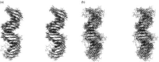
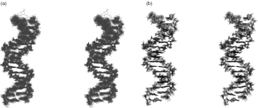
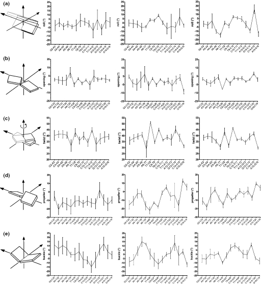
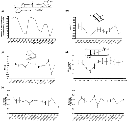
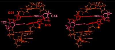
The integration of the human immunodeficiency virus type 1 DNA into the host cell genome is catalysed by the viral integrase (IN). The reaction consists of a 3'-processing [dinucleotide released from each 3' end of the viral long terminal repeat (LTR)] followed by a strand transfer (insertion of the viral genome into the human chromosome). A 17 base pair oligonucleotide d(GGAAAATCTCTAGCAGT), d(ACTGCTAGAGATTTTCC) reproducing the U5-LTR extremity of viral DNA that contains the IN attachment site was analysed by NMR using the classical NOEs and scalar coupling constants in conjunction with a small set of residual dipolar coupling constants (RDCs) measured at the 13C/15N natural abundance. The combination of these two types of parameters in calculations significantly improved the DNA structure determination. The well-known features of A-tracts were clearly identified by RDCs in the first part of the molecule. The binding/cleavage site at the viral DNA end is distinguishable by a loss of regular base stacking and a distorted minor groove that can aid its specific recognition by IN.


Figures

Figure 1
Figure 1
Stereo views of the nine lowest-energy structures of U5-term generated from classical scalar coupling and NOE restraints. These were optimized by relaxation matrix refinement using: (a) uncertainties of ±20%; (b) very weak, weak, medium and strong distance restraint categories.
Figure 2
Figure 2
Stereo views of the U5-term structures corresponding to: (a) 36 RDC-structures, i.e. four families of nine best structures each family being generated from a different value of alignment tensor (see Table 2); (b) nine lowest-energy structures generated from classical NOE and scalar coupling restraints combined to RDC restraints (Da = −22 Hz; R = 0.1).
Figure 3
Figure 3
Structural parameters of U5-term. Profiles of: (a) inter base (intrastrand) rolls in the upper strand; (b) global base–base openings; (c) inter base twists in the upper strand; (d) global base–base propellers; (e) global base–base buckles. Values were extracted from outputs of CURVES (39) for: in left, a set of nine structures generated from NOE restraints classified into very weak, weak, medium and strong distance categories; in middle, a set of nine structures generated from NOE restraints with uncertainties of ±20%; and in right, the 36 RDC structures (see legend in Figure 2).
Figure 4
Figure 4
(a) Profiles of the local number of B-DNA structures (Zp value inferior to 0.5 Å) among the 36 RDC structures [calculated with 3DNA (42)]. A schematic definition of Zp is given in insert (41); (b) Profile of the global base pair axis inclinations of the 36 RDC structures (see legends in Figure 3 and Table 2), values being calculated with CURVES (39); (c) Profiles of local inter base (intrastrand) tilts in the upper strand of the 36 RDC structures (see legend in Figure 3), values being calculated with CURVES (39); (d) Profile of the average interstrand phosphorus–phosphorus (Pm–Pn+3) distances (illustrating the minor groove width) from the 36 RDC structures (see legend in Figure 3), calculated with 3DNA (42); (e) Profiles of intrastrand phosphate–phosphate Pi–Pi+1 distances along the upper strand (left) and the lower strand (right).
Figure 5
Figure 5
A stereo view of the average structure of the five outermost base pairs obtained from the 36 RDC structures of U5-term (see legend in Figure 2) featuring the particular interstrand stacking of purines A15 and G21 in red thick lines and the phosphorus of the scissile bond in CPK.

Similar articles

Cited by

References

    1. Hazuda D.J., Felock P., Witmer M., Wolfe A., Stillmock K., Grobler J.A., Espeseth A., Gabryelski L., Schleif W., Blau C., Miller M.D. Inhibitors of strand transfer that prevent integration and inhibit HIV-1 replication in cells. Science. 2000;287:646–650. - PubMed
    1. Pommier Y., Neamati N. Inhibitors of human immunodeficiency virus integrase. Adv. Virus Res. 1999;52:427–458. - PubMed
    1. Esposito D., Craigie R. HIV integrase structure and function. Adv. Virus Res. 1999;52:319–333. - PubMed
    1. Katzman M., Katz R.A. Substrate recognition by retroviral integrases. Adv. Virus Res. 1999;52:371–395. - PubMed
    1. Gerton J.L., Brown P.O. The core domain of HIV-1 integrase recognizes key features of its DNA substrates. J. Biol. Chem. 1997;272:25809–25815. - PubMed

Publication types