. 2005 Apr 6;33(6):1982-92.
doi: 10.1093/nar/gki348. Print 2005.

Cyclic changes in the affinity of protein-DNA interactions drive the progression and regulate the outcome of the Tn10 transposition reaction

Affiliations

Cyclic changes in the affinity of protein-DNA interactions drive the progression and regulate the outcome of the Tn10 transposition reaction

Danxu Liu et al. Nucleic Acids Res. .

Abstract

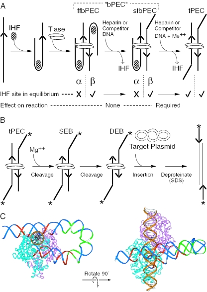
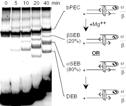
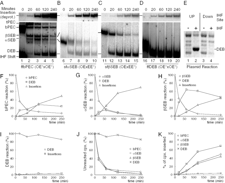
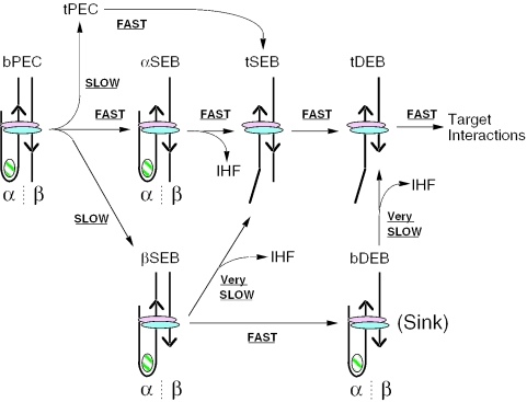
The Tn10 transpososome is a DNA processing machine in which two transposon ends, a transposase dimer and the host protein integration host factor (IHF), are united in an asymmetrical complex. The transitions that occur during one transposition cycle are not limited to chemical cleavage events at the transposon ends, but also involve a reorganization of the protein and DNA components. Here, we demonstrate multiple pathways for Tn10 transposition. We show that one series of events is favored over all others and involves cyclic changes in the affinity of IHF for its binding site. During transpososome assembly, IHF is bound with high affinity. However, the affinity for IHF drops dramatically after cleavage of the first transposon end, leading to IHF ejection and unfolding of the complex. The ejection of IHF promotes cleavage of the second end, which is followed by restoration of the high affinity state which in turn regulates target interactions.


Figures

Figure 1
Figure 1
Assembly of the Tn10 transpososome and the chemical steps of the reaction. (A) Assembly and unfolding of the synaptic complex. Synapsis of two IHF-bound transposon arms by transposase produces an ffbPEC. Treatment with competitor DNA or heparin strips IHF from the β side of the complex to produce sfbPEC. IHF remains bound to the α side of the complex, presumably due to the subterminal transposase contacts with the transposon end. Divalent metal ion unlocks the IHF binding site on the α side of the complex. Dissociation of IHF produces the tPEC. Hatched oval, IHF; open ovals, transposase; and arrowhead, transposon end. In the tPEC, unoccupied IHF binding sites are indicated by a kink in the transposon end. (B) The chemical steps of the transposition reaction are illustrated using the tPEC as the starting point. In the presence of Mg++, the flanking DNA is cleaved to produce a SEB, followed by a DEB. Non-covalent interactions with a target site are followed by the strand transfer step that produces an insertion product. Asterisk, location of the 32P-label on the transposon end; other elements are as described in (A). (C) The Tn10 transpososome was modeled by superimposing the DNA from the IHF co-crystal structure onto the structure of the Tn5 transpososome (28,32,44,45). Superimposition of the IHF-folded DNA was achieved by minimizing the RMS difference in the position of the equivalent atoms in the Tn5 DNA. One transposon end, IHF and flanking DNA have been omitted for clarity. A section of B DNA (gold) has been docked in the target binding groove to illustrate its spatial relationship to the IHF-folded transposon arm. Regions of transposase and IHF mediated hydroxyl radical protection are shown in red and green, respectively. Every tenth nucleotide on the transferred strand is shown in white. The transposon end is seen embedded in the turquoise monomer of transposase. The subterminal transposase contacts are located on the top of the structure illustrated on the left.
Figure 2
Figure 2
Slow unfolding of the bPEC and DEB complexes. (A) bPEC was assembled and treated with heparin and Ca++ to initiate unfolding. The kinetics of unfolding was monitored using the EMSA. Autoradiograms are shown. Left panel: the transpososome contains two identical outside ends of Tn10. In this and most other experiments, the IHF concentration was limited so that just over 50% of the transposon end DNA fragments are shifted (lanes 2 and 3). The bPEC therefore represents a mixture of ffbPEC and sfbPEC which comigrate in the gel (see Figure 1A). Right panel: the sfbPEC was prepared directly by mixing a radioactively labeled even-end with an unlabeled outside end. The even-end is analogous to the inside end of IS10 that lacks an IHF binding site. The even-end will not form complex without an outside end partner. Since the label is present on the even-end, only mixed complexes are detected. Also, since the even-end lacks an IHF binding site, no IHF-shifted DNA is observed. In the left panel, the outside end was prepared by digesting pRC98 with XbaI+HincII (85 bp transposon arm/62 bp flanking DNA). In the right panel, the outside end was prepared by digesting pRC98 with AccI+SacII (84 bp transposon arm/75 bp flanking DNA). The even-end DNA fragment was prepared by digesting pRC100 with AccI+BamHI (73 bp transposon arm/39 bp flanking DNA). Details of the additions made to the reactions, indicated above the gel, are given in Materials and Methods. OE, outside end; EE, even-end; other details are as given in Figure 1. (B) The DEB complexes were prepared as described in (A) using precleaved transposon ends. The outside end was prepared by digesting pRC35 with BstEII+PvuII (85 bp transposon arm). The even-end DNA fragment was prepared by digesting pRC99 with AccI+PvuII (73 bp transposon arm). Other details are as given in (A).
Figure 3
Figure 3
Identification of two isomers of the SEB complex. (A) SEB complex was assembled by mixing a radioactively labeled uncleaved outside end with an excess of unlabeled precleaved outside end. This biases the reaction strongly in favor of the assembly of mixed complexes. The uncleaved outside end was prepared by digesting pRC167 with PstI+XhoI (96 bp transposon arm/77 bp flanking DNA). The precleaved outside end was prepared by digesting pRC35 with PvuII+BstEII (87 bp transposon arm). sfαSEB, semi-folded α-single-end-break; sfβSEB, semi-folded β-single-end-break. Other details are as given in Figures 1 and 2. (B) The sfαSEB and sfβSEB were footprinted with hydroxyl radicals. Briefly, complexes were assembled, treated in solution with hydroxyl radicals and separated using the EMSA. The complexes were recovered from the gel and the footprints were displayed on a DNA sequencing gel. Dark and light shaded boxes represent the transposase and IHF footprints as described previously (32). The large arrowhead indicates the location of the transposon end. The number of base pair inside the transposon is indicated.
Figure 4
Figure 4
Kinetics and requirements for unfolding of the SEB isomers. Mixed complexes were assembled using the transposon ends illustrated below each panel. There was a 4-fold molar excess of the outside end present. The outside end was prepared by digesting pRC98 with AccI+ScaI (84 bp transposon arm/75 bp flanking DNA). The precleaved outside end was prepared by digesting pRC35 with BstEII+PvuII (85 bp transposon arm). The even-end DNA fragment was prepared by digesting pRC100 with AccI+BamHI (73 bp transposon arm/39 bp flanking DNA). The precleaved even-end was prepared by digesting pRC99 with AccI+PvuII (73 bp transposon arm). Other details are as given in Figure 2.
Figure 5
Figure 5
The cleavage step is biased toward the αSEB isomer. The standard complex assembly reaction was scaled up and initiated at time zero by the addition of Mg++. Aliquots were removed at the indicated times and loaded directly onto the gel that was under electrical tension. The outside end and even-end DNA fragments were prepared by digesting pRC167 and pRC100 with PstI+XhoI and HindIII+SpeI, respectively. To preclude any potential influence on the rate of cleavage by the sequence of bases flanking the transposon, the flanking DNA on each fragment is isogenic. The outside and even-ends are also isogenic out to bp 19 of the transposon end. Other details are as given in Figure 2.
Figure 6
Figure 6
Insertion kinetics of the different complexes. (AD) and (FK) The standard 20 μl reaction was scaled up to 200 μl and initiated at time zero by the addition of Mg++ in a mixture with 10 μg of pBluescript as target DNA. Aliquots (40 μl) were withdrawn at the indicated time and stopped by the addition of EDTA. Half of the reaction was deproteinated by SDS treatment to allow detection of the large insertion product. For each type of complex, the native and deproteinated aliquots (20 μl) were electrophoresed on the same gel so that the quantification of the various complexes and products was exactly comparable and did not require normalization. The sequence of bases flanking the transposon ends on each fragment was isogenic to preclude any potential effect on the rate of cleavage. bPEC panel; the outside end was prepared by digesting pRC98 with AccI+SacII (84 bp transposon arm/75 bp flanking DNA). αSEB panel: the precleaved outside end was prepared by digesting pRC35 with BstEII+PvuII (85 bp transposon arm); the uncleaved even-end DNA fragment was prepared by digesting pRC100 with AccI+BamHI (73 bp transposon arm/39 bp flanking DNA). βSEB panel: the uncleaved outside end was prepared by digesting pRC98 with AccI+SacII (84 bp transposon arm/75 bp flanking DNA); the precleaved even-end was prepared by digesting pRC99 with AccI+PvuII (73 bp transposon arm). DEB panel: the precleaved outside end was prepared by digesting pRC35 with BstEII+PvuII (85 bp transposon arm). (E) Transposition reactions (200 μl) contained 1 nM plasmid substrate encoding a mini-Tn10 transposon, 6 nM transposase and 35 nM IHF. The reactions were incubated at 30°C for 3 h, concentrated by ethanol precipitation and analyzed on a TBE-buffered 1.1% agarose gel (30,39). Excision of the transposon segment from the supercoiled plasmid substrate produces the DEB product (referred to in previous publications as the ‘excised transposon fragment’ or ETF). Auto-integration of the DEB product produces a topologically complex set of knot and catenane products (39). Apart from the DEB product, these are the only products present in the section of the gel shown. The IHF-up and IHF-down mutations were encoded on plasmids pNK2588 and pNK2590, respectively. IHF-up (single point mutation) and IHF-down (triple point mutation) move the IHF binding site closer to or further away from the IHF consensus binding site as described in Figure 1 of ref. (46). A reverse contrast photograph of an ethidium bromide stained agarose gel is shown.
Figure 7
Figure 7
Multiple pathways for the unfolding and cleavage of the Tn10 transpososome. The model summarizes the potential of the transpososome to unfold at different stages of the cleavage reaction. At the start of the reaction, the bPEC can follow one of the three pathways. The least productive is via the βSEB intermediate. Transpososomes that arrive at the DEB stage of the reaction with IHF still associated, or reacquire IHF at this stage of the reaction, unfold and perform the strand transfer step very slowly.

Similar articles

Cited by

References

    1. Katinka M.D., Duprat S., Cornillot E., Metenier G., Thomarat F., Prensier G., Barbe V., Peyretaillade E., Brottier P., Wincker P., et al. Genome sequence and gene compaction of the eukaryote parasite Encephalitozoon cuniculi. Nature. 2001;414:450–453. - PubMed
    1. Mahillon J., Chandler M. Insertion sequences. Microbiol. Mol. Biol. Rev. 1998;62:725–774. - PMC - PubMed
    1. Davis B.M., Waldor M.K. Mobile genetic elements and bacterial pathogenesis. In: Craig N.L., Craigie R., Gellert M., Lambowitz A.M., editors. Mobile DNA II. Washington, DC: American Society for Microbiology; 2002. pp. 1040–1059.
    1. Campbell A. Eubacterial genomes. In: Craig N.L., Craigie R., Gellert M., Lambowitz A.M., editors. Mobile DNA II. Washington, DC: American Society for Microbiology; 2002. pp. 1024–1039.
    1. Chalmers R., Blot M. Insertion sequences and transposons. In: Charlebois R.L., editor. Organization of the Prokaryotic Genome. Washington, DC: American Society for Microbiology; 1999. pp. 151–169.

Publication types