. 2005 Apr 6;33(6):1993-2002.
doi: 10.1093/nar/gki347. Print 2005.

Functional roles of 3'-terminal structures of template RNA during in vivo retrotransposition of non-LTR retrotransposon, R1Bm

Affiliations

Functional roles of 3'-terminal structures of template RNA during in vivo retrotransposition of non-LTR retrotransposon, R1Bm

Tomohiro Anzai et al. Nucleic Acids Res. .

Abstract

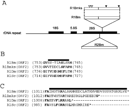
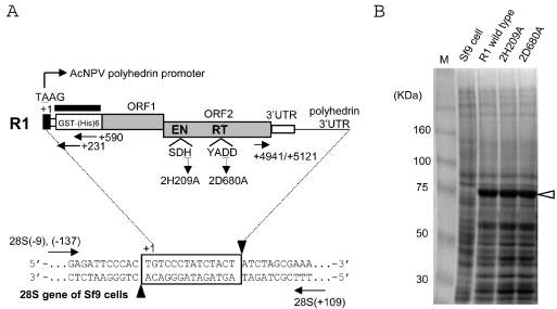
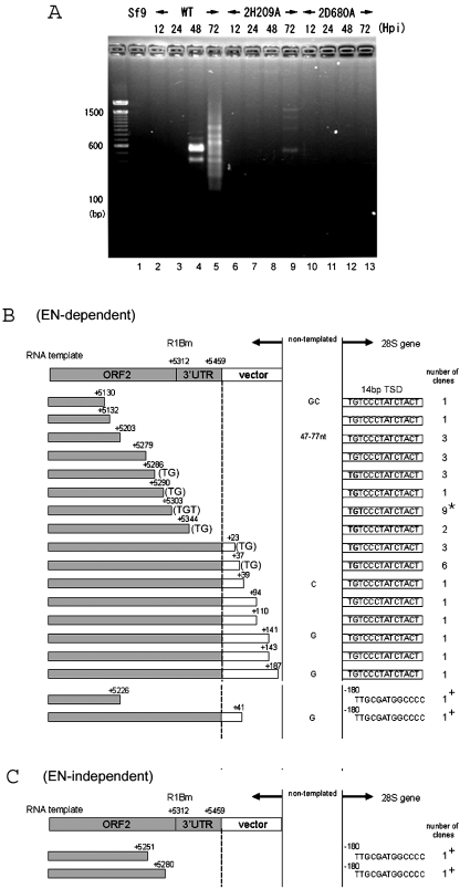
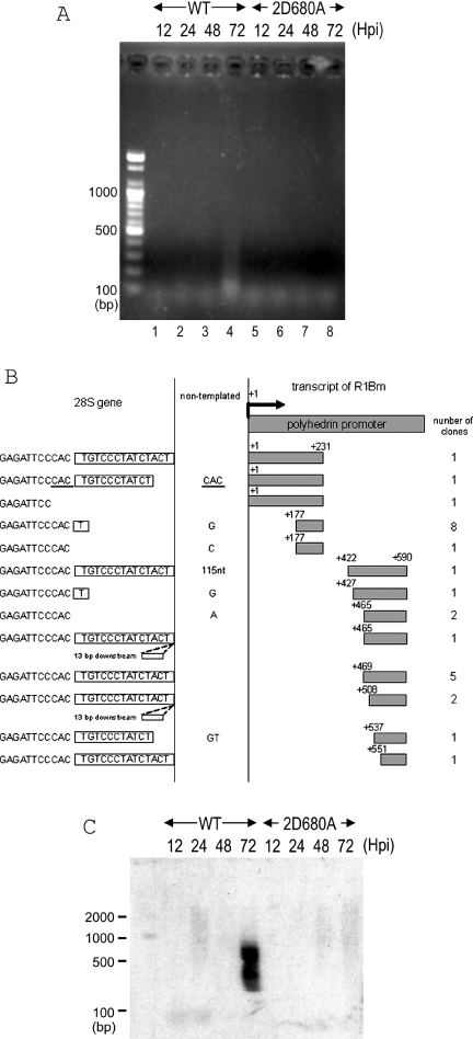
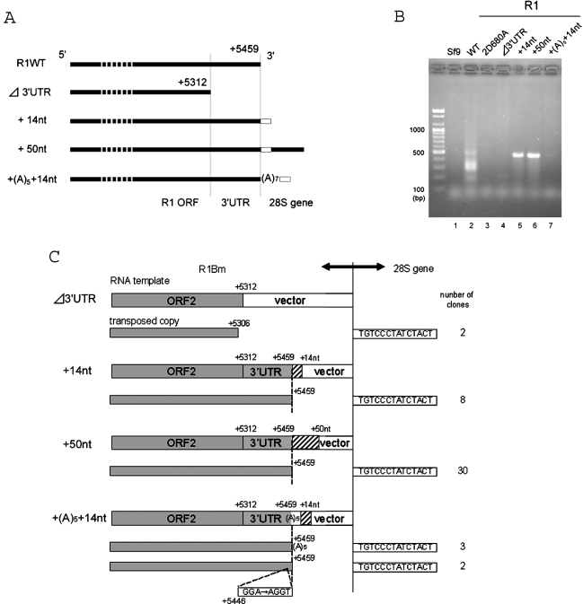
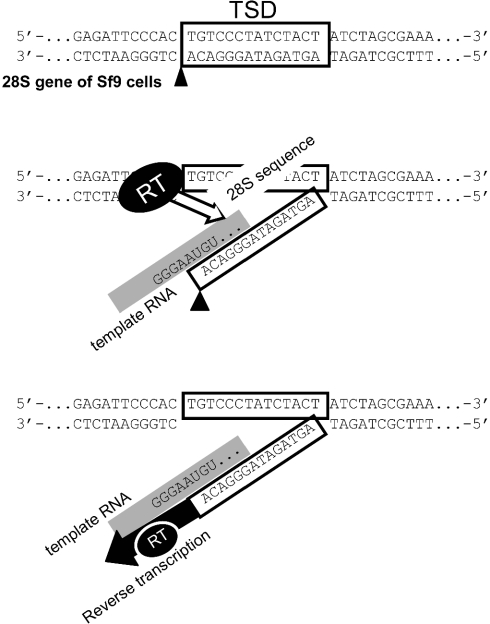
R1Bm is a non-LTR retrotransposon found specifically within 28S rRNA genes of the silkworm. Different from other non-LTR retrotransposons encoding two open reading frames (ORFs), R1Bm structurally lacks a poly (A) tract at its 3' end. To study how R1Bm initiates reverse transcription from the poly (A)-less template RNA, we established an in vivo retrotransposition system using recombinant baculovirus, and characterized retrotransposition activities of R1Bm. Target-primed reverse transcription (TPRT) of R1Bm occurred from the cleavage site generated by endonuclease (EN). The 147 bp of 3'-untranslated region (3'UTR) was essential for efficient retrotransposition of R1Bm. Even using the complete R1Bm element, however, reverse transcription started from various sites of the template RNA mostly with 5'-UG-3' or 5'-UGU-3' at their 3' ends, which are presumably base-paired with 3' end of the EN-digested 28S rDNA target sequence, 5'-AGTAGATAGGGACA-3'. When the downstream sequence of 28S rDNA target was added to the 3' end of R1 unit, reverse transcription started exactly from the 3' end of 3'UTR and retrotransposition efficiency increased. These results indicate that 3'-terminal structure of template RNA including read-through region interacts with its target rDNA sequences of R1Bm, which plays important roles in initial process of TPRT in vivo.


Figures

Figure 1
Figure 1
Identification of a new subtype of the R1 element from the silkworm, Bombyx mori. (A) Schematic representation of 28S gene-specific LINEs. Shown in the middle is a diagram of the rDNA unit of Bombyx mori. The newly identified R1 clone (named R1Bmks) is integrated into the sequence 74 bp downstream of the R2 insertion site. Open reading frames (ORFs) are depicted by open boxes. The positions of non-synonymous mutations in the ORF1 and nucleotide insertions in the ORF2 (see B and C) are shown by white and black arrowheads, respectively. (B and C) Altered amino acid sequence of R1Bmks in the ORF2. Insertion of 3 nt within the RT domain results in alteration of 10 amino acids (indicated by bold line), compared with R1Bm (B). Addition of a single nucleotide in R1Bmks alters the amino acid sequences and made the length of ORF2 shorter by 12 amino acids, compared with R1Bm (C). The amino acids conserved between more than two elements are indicated by bold letters. Putative translational stop was indicated by asterisks. R1Bm clones are aligned with R1Dm (X51968; accession number) from D.melanogaster; R1Sc (L00945) from Sciara coprophila.
Figure 2
Figure 2
In vivo retrotransposition assay for R1 elements using recombinant baculoviruses. (A) Diagram of the PCR assay for retrotransposition. Shown at the top of the figure is a diagram of the R1 element expressed from AcNPV. The 5′UTR sequence derived from the polyhedrin promoter and ORF1/ORF2 of R1 are shaded in black and gray, respectively. Nucleotide position is numbered with the transcription initiation site (A of TAAG) defined as +1. EN and RT denote the endonuclease and reverse transcriptase domains, respectively. The amino acid position of each mis-sense mutant is also shown; the 2H209A mutant represents the substitution of the 209th histidine (H) in the ORF2 for alanine (A), and the 2D680A mutant represents the substitution of the 680th aspartic acid (D) in the ORF2 for alanine (A). Shown at the bottom is a diagram of an rDNA unit showing the location of R1 insertion; 14 bp of the target site duplication (TSD) created upon R1 insertion is boxed. The putative cleavage sites by R1-EN are indicated by black arrowheads. Arrows represent the primer set to amplify the 5′ and 3′ junctions between the R1 sequence and the 28S sequence. The thick bar above the GST region which is fused to the R1 ORF1 indicates the probe used for Southern hybridization in Figure 4. (B) Expression of R1 proteins with a baculovirus-based expression system. The total proteins were extracted from Sf9 cells infected with recombinant viruses shown above and run on SDS–PAGE; the predicted molecular weight for R1 GST/(His)6/ORF1 was 78 170. Lane M; size markers.
Figure 3
Figure 3
3′-junction analysis for retrotransposed R1 elements. (A) PCR amplification of the 3′ boundaries between the transposed R1 and the 28S rDNA gene. Sf9 genomic DNAs were extracted 12, 24, 48, and 72 h post-infection (h.p.i.) with AcNPV expressing wild-type R1, 2H209A (EN-deficient mutant) and 2D680A (RT-deficient mutant). The purified DNA was used as template for PCR amplification with a pair of primers, +4941 and 28S(+109) (Figure 2A). The PCR products were subjected to 2.5% agarose electrophoresis and stained with ethidium bromide. The molecular size marker is loaded alongside and each size in base pair (bp) was shown on the left of the picture. (B and C) 72 h.p.i. 3′ junction clones obtained with wild-type R1 (B) and endonuclease-deficient R1 (2H209A) (C). Shown at the top of each figure is a diagram of the 3′ end structure of the construct. Sequences derived from the R1Bm and the pAcGHLTB vectors are indicated by shaded and open boxes, respectively. The initiation sites for reverse transcription (left of the dotted vertical lines) are indicated by nucleotide numbers. The target DNA regions are shown on the right of the dotted vertical lines. Extra nucleotides at the junction that are not derived from either the 28S gene or the R1 construct are given between the two vertical lines (non-templated). Boxes to the right of the vertical lines represent the 14 bp of TSD. The number of clones containing each insertion type is indicated in the right-most column and the most major type is indicated by an asterisk. The TGT or TG sequences on the 3′ end of the R1 template that can base-pair with the target DNA are also indicated (Figure 6). +, insertions into the site 180 bp upstream of TSD observed for wild-type and endonuclease-deficient R1.
Figure 4
Figure 4
5′-junction analysis for retrotransposed R1 elements. (A) PCR amplification of the boundaries of the transposed R1 5′ ends with the 28S gene. The DNA extraction and PCR analyses were basically same as in Figure 3. A pair of primers, +590 and 28S(−62) was used for amplifying the 5′ junctions. 2D680A, RT-deficient mutant (Figure 2A). (B) 72 h.p.i. 5′ junction obtained with wild-type R1. Shown at the top of the figure is a diagram of the 5′ end structure of R1WT-pAcGHLT. Sequences derived from the pAcGHLTB vector are indicated by shaded boxes. The numbers on the left and the right of the shaded boxes correspond to the nucleotide position numbered with the transcription initiation site. The gray boxes on the right are cDNA and the open boxes on the left are the 28S rDNA regions. Extra nucleotides at the junction that are not derived from either the 28S gene or the R1 construct are given between two vertical lines. Boxes to the left of the vertical lines represent the 14 bp of TSD. Duplicated nucleotides are underlined. Two of the insertions contained 13 bp downstream sequences at the insertion site. The number of clones containing each insertion type is indicated in the right-most column. The top five sequences are from PCR with primer +231, and the bottom eight sequences are from PCR with primer +590. (C) Southern hybridization of the retrotransposed R1 5′ junction PCR. The PCR reaction was conducted by primers +590 and 28S (−137). The PCR products were transferred to a nylon membrane and hybridized with the GST probe (Figure 2A). The DNA size marker was electrophoresed simultaneously and shown on the left.
Figure 5
Figure 5
Effects of downstream sequences on the R1 retrotransposition. (A) Diagram of the R1 constructs with various 3′ end structures; 14 and 50 nt of the downstream 28S gene are added to the R1WT construct. The 14 bp sequence of TSD is indicated by an open box. Note that the 3′ termini of each construct is followed by the AcNPV-derived polyhedrin 3′UTR. Transcription start, +1. (B) PCR amplification of the 3′ boundaries between the transposed R1 copies and the 28S gene. DNA extraction and PCR reaction are basically same as in Figure 3. The primer set used for PCR was +5121 and 28S(+109). 2D680A, RT-deficient mutant. (C) 72 h.p.i. 3′ junctions obtained with various R1 construct in (A). Shown at the top of each figure is a diagram of the 3′ end structure of the constructs. Sequences derived from R1Bm and additional 28S downstream sequences are indicated by shaded and hatched boxes, respectively. The vertical dotted lines represent the boundary between the cDNA region and the target DNA region. The initiation site for reverse transcription (left of the dotted vertical lines) is indicated by the numbers. Boxes to the right of the vertical lines represent the 14 bp of TSD. The number of clones containing each insertion type is indicated in the right-most column. Two clones amplified from the + (A)5 + 14 nt construct contained mutations at the 3′ end of 3′UTR (G of GAA corresponds to the position +5446).
Figure 6
Figure 6
Schematic representation of initial process of TPRT in the R1 element. (Top) First, R1-EN cleaves the non-coding bottom strand of 28S rDNA in the A–C junction. Boxes represent the 14 bp of TSD. (Middle) Then, the target DNA is partially denatured, allowing the UGU on the RNA template to base-pair with the loose target DNA. The template RNA is indicated by a gray line. In this model, the read-through 28S rRNA sequence is base-paired with the DNA target in longer region. During this process, RT of R1Bm may recognize the 3′UTR and base-paired region (open arrow), and place the RNA template at the accurate position for initiation of reverse transcription. (Bottom) Next, reverse transcription starts from the position next to the UGU sequence of template RNA, using the 3′-OH of A residue as primer.

Similar articles

Cited by

References

    1. Arkhipova I., Meselsonm M. Transposable elements in sexual and ancient asexual taxa. Proc. Natl Acad. Sci. USA. 2000;97:14473–14477. - PMC - PubMed
    1. Lander E.S., Linton L.M., Birren B., Nusbaum C., Zody M.C., Baldwin J., Devon K., Dewar K., Doyle M., FitzHugh W., et al. Initial sequencing and analysis of the human genome. Nature. 2001;409:860–921. - PubMed
    1. Moran J.V., DeBerardinis R.J., Kazazian H.H., Jr Exon shuffling by L1 retrotransposition. Science. 1999;283:1530–1534. - PubMed
    1. Gilbert N., Lutz-Prigge S., Moran J.V. Genomic deletions created upon LINE-1 retrotransposition. Cell. 2002;110:315–325. - PubMed
    1. Symer D.E., Connelly C., Szak S.T., Caputo E.M., Cost G.J., Parmigiani G., Boeke J.D. Human L1 retrotransposition is associated with genetic instability in vivo. Cell. 2002;110:327–338. - PubMed

Publication types