. 2005 Jun;16(6):2848-61.
doi: 10.1091/mbc.e05-01-0067. Epub 2005 Apr 6.

The ATP-dependent membrane localization of protein kinase Calpha is regulated by Ca2+ influx and phosphatidylinositol 4,5-bisphosphate in differentiated PC12 cells

Affiliations

The ATP-dependent membrane localization of protein kinase Calpha is regulated by Ca2+ influx and phosphatidylinositol 4,5-bisphosphate in differentiated PC12 cells

Consuelo Marín-Vicente et al. Mol Biol Cell. 2005 Jun.

Abstract

Signal transduction through protein kinase Cs (PKCs) strongly depends on their subcellular localization. Here, we investigate the molecular determinants of PKCalpha localization by using a model system of neural growth factor (NGF)-differentiated pheochromocytoma (PC12) cells and extracellular stimulation with ATP. Strikingly, the Ca2+ influx, initiated by the ATP stimulation of P2X receptors, rather than the Ca2+ released from the intracellular stores, was the driving force behind the translocation of PKCalpha to the plasma membrane. Furthermore, the localization process depended on two regions of the C2 domain: the Ca2+-binding region and the lysine-rich cluster, which bind Ca2+ and phosphatidylinositol 4,5-bisphosphate [PtdIns(4,5)P2], respectively. It was demonstrated that diacylglycerol was not involved in the localization of PKCalpha through its C1 domain, and in lieu, the presence of PtdIns(4,5)P2 increased the permanence of PKCalpha in the plasma membrane. Finally, it also was shown that ATP cooperated with NGF during the differentiation process of PC12 cells by increasing the length of the neurites, an effect that was inhibited when the cells were incubated in the presence of a specific inhibitor of PKCalpha, suggesting a possible role for this isoenzyme in the neural differentiation process. Overall, these results show a novel mechanism of PKCalpha activation in differentiated PC12 cells, where Ca2+ influx, together with the endogenous PtdIns(4,5)P2, anchor PKCalpha to the plasma membrane through two distinct motifs of its C2 domain, leading to enzyme activation.


Figures

Figure 1.
Figure 1.
Protein kinase Cα translocates to the plasma membrane of dPC12 cells via ATP stimulation. (A) Confocal images of dPC12 cells expressing protein kinase Cα-EGFP and stimulated with 100 μM ATP. The frames shown in the figure correspond to 0 and 20 s after ATP stimulation. Protein kinase Cα-EGFP fluorescence intensity was measured in the cytosol and in the plasma membrane in every frame of the time series (1 frame every 10 s). (B) The protein localization was measured by a line profile (pixel density) traced in each frame as indicated in Materials and Methods procedures section and analyzed with the program ImageJ NIH. The resulting net change in protein kinase Cα localization is expressed as the Imb-Icyt/Imb ratio (R) and is represented versus time (n = 24 cells). The dotted line represent an average of the time course of [Ca2+]i after ATP stimulation, which has been taken from part C of this figure and inserted here to facilitate the interpretation of the data. (C) Time course of [Ca2+]i fluctuations were monitored with Fura Red, the figure shows different profiles representative of the wide range of the [Ca2+]i observed (n = 125 cells). It is important to note that all of them correlated with protein kinase Cα translocation to the plasma membrane after ATP stimulation.
Figure 2.
Figure 2.
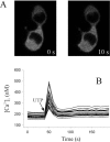
UTP stimulation does not induce protein kinase Cα localization in the plasma membrane of dPC12 cells. (A) Confocal images corresponding to dPC12 cells treated with 100 μM UTP at 0 and 10 s after stimulation. (B) Time course of [Ca2+]i variations in dPC12 cells loaded with Fura Red-AM ester and stimulated with 100 μM UTP. The profiles represented in the figure are representative of the cells analyzed (n = 32) and none of them correlated with protein kinase Cα translocation to the plasma membrane after UTP stimulation.
Figure 3.
Figure 3.
ATP evoked Ca2+ influx is necessary to drive protein kinase Cα to the plasma membrane. Time course of [Ca2+]i variations of dPC12 cells loaded with Fura Red-AM ester and stimulated with 100 μM ATP. The cells were incubated with extracellular medium containing Ca2+ free HBS (from 0 to 150 s) and stimulated with ATP at the time indicated by the arrow. At 150 s of the recording, the cells were washed with 4 ml of HBS containing 3 mM CaCl2 and stimulated again with ATP, which now induced a Ca2+ peak, ranging from 1.3 to 4 μM. Confocal images (a–c) of dPC12 cells transfected with protein kinase Cα-EGFP, taken at the times indicated by the corresponding letters a–c in the [Ca2+]i time profile.
Figure 4.
Figure 4.
Threshold value of [Ca2+]i for translocation of protein kinase Cα in dPC12 cells. The cells were loaded with Fura Red-AM ester and preincubated with HBS containing different concentrations of extracellular Ca2+. (A) Time course of [Ca2+]i variations when the cells (n = 32) were incubated with HBS containing 750 μM CaCl2 and stimulated with ATP. The panels show dPC12 cells transfected with protein kinase Cα-EGFP in a representative experiment in resting conditions (a) and 20 s after ATP stimulation (b). (B) Time course of [Ca2+]i variations when the cells (n = 30) were incubated with HBS containing 500 μM CaCl2 and stimulated with ATP. No protein kinase Cα-EGFP membrane localization was observed in these conditions. Note that the [Ca2+]i is a transient peak that only reached 830 nM, which seems insufficient to promote translocation of protein kinase Cα to the plasma membrane. The concentration of free Ca2+ was estimated from total concentration of Ca2+and EGTA by using computer software developed by Fabiato (1988).
Figure 5.
Figure 5.
The Ca2+-binding region of the C2 domain is a key motif in the membrane localization of protein kinase Cα. Confocal images of dPC12 cells expressing protein kinase Cα-D187N/D246N/D248N-EGFP mutant before (a) and 20 s (b) after ATP stimulation are shown (n = 21 cells). The protein kinase Cα mutant is cytosolic in unstimulated cells and does not translocate to the plasma membrane after the addition of ATP in any of the frames analyzed. Confocal images were recorded every 10 s for 10 min.
Figure 6.
Figure 6.
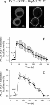
Inhibition of PI-PLC increases the localization time of protein kinase Cα in the plasma membrane. (A) dPC12 cells expressing protein kinase Cα-EGFP were pretreated with 10 μM U73122 for 20 min before ATP stimulation. Translocation of the protein to the plasma membrane started 10 s after ATP addition. (B) Time profile of the effect of U73122 on the protein kinase Cα-EGFP membrane localization that is expressed as relative plasma membrane translocation, R (n = 15 cells). (C) Time profile of the effect of preincubation for 20 min with 50 mM 1-butanol on the protein kinase Cα-EGFP membrane localization, R (n = 21 cells).
Figure 7.
Figure 7.
Neither ATP nor UTP stimulation induces the ECFP-C1a domain translocation to the plasma membrane. (A) The C1a domain of protein kinase Cα was fused to ECFP and expressed in dPC12 cells after ATP (top) or UTP (bottom) stimulation. Confocal images were collected every 10 s for 10 min. Observe that the domain localized both in the cytosol and in the cells nucleus, as described previously by Oancea and Meyer (1998). (B) To confirm the functionality of the cloned C1a domain, the transfected cells were incubated with increasing concentrations of extracellular DiC8 and the maximal relative membrane localization (R) was calculated for each of the DiC8 concentrations used. The apparent half-maximal [DiC8] was calculated graphically.
Figure 8.
Figure 8.
The membrane localization of protein kinase Cα after ATP stimulation induces the dissociation of the PH domain of PLCδ. (A) Ca2+ influx driven by ATP stimulation does not induce ECFP-PH-PLCδ membrane dissociation. Confocal images of dPC12 cells expressing ECFP-PH-PLCδ domain (0 s), which localizes heterogeneously in areas of the plasma membrane of dPC12 cells. The PH-PLCδ domain did not exhibit a transient dissociation from the plasma membrane after ATP stimulation (30 s), as might be expected if this stimulation induced the hydrolysis of the endogenous PtdIns(4,5)P2. (B) dPC12 cells were cotransfected with ECFP-PH-PLCδ (left) and protein kinase Cα-EYFP (right) constructs. Confocal images of simultaneous double detection of ECFP and EYFP were obtained by excitation at 458 and 514 nm, a double dichroic filter DD458/514 and emission wavelengths recorded in two independent channels at 470–490 and 540–582 nm. It was tested that under these conditions there was no bleed-through of the CFP into the YFP emission channels. A series of 30–60 confocal images were recorded for each experiment at time intervals of 10 s. Top, localization of each construct in resting conditions. Bottom, localization of each construct 30 s after the ATP stimulation that induced protein kinase Cα translocation to the plasma membrane and a simultaneous dissociation of the PH domain. (C) Time course of the relative membrane localization of both constructs: ECFP-PH-PLCδ (○) and protein kinase Cα-EYFP (▪). The maximal relative membrane localization was expressed as R and calculated as described in Materials and Methods.
Figure 9.
Figure 9.
ATP stimulation induces a transient membrane localization of a lysine-rich cluster protein kinase Cα mutant. (A) dPC12 cells were cotranfected with the ECFP-PH-PLCδ domain, a PtdIns(4,5)P2 biosensor (left) and with the protein kinase CαK209A/K211A-EYFP mutant (right). The images represent cells in resting conditions (top) and 30 s after stimulation with 100 μM ATP (bottom). Partial plasma membrane localization of the protein kinase Cα mutant was observed, which correlated with a lack of dissociation of the PH domain from the plasma membrane. Confocal images were collected in a similar way to that used in the experiment depicted in Figure 8. (B) Time course of the relative membrane localization of protein kinase CαK209A/K211A-EYFP mutant (▪). The maximal relative membrane localization was expressed as R and calculated as described in Materials and Methods.
Figure 10.
Figure 10.
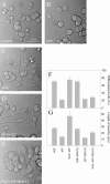
Extracellular ATP induces neurite growth through protein kinase Cα signaling. PC12 cells were differentiated for 48 h in a medium containing DMEM supplemented with 5% heat inactivated horse serum, 2.5% FBS in the presence of either 100 μM ATP (B) or 100 ng/ml NGF (C). (D) Image representative of a PC12 cell culture differentiated with 100 μM ATP and 100 ng/ml NGF simultaneously. (E) PC12 cells differentiated in the presence of ATP, NGF, and 20 nM BIM XI, which is a specific protein kinase Cα inhibitor. All the images correspond to phase contrast micrographs taken after 48 h. Bar, 40 μm. The response of PC12 cells to the different incubation conditions was quantified by two criteria: number of neurite-bearing cells (F) and length of neurites (G), at least 100 cells from 10 randomly chosen fields were scored. Control cells (A) were kept for 48 h under the same conditions except that NGF or ATP were not added to the differentiation media.
Figure 11.
Figure 11.
Model for plasma membrane translocation of protein kinase Cα in dPC12 cells stimulated with ATP. The ATP stimulation of dPC12 cells induces a Ca2+ influx through P2X receptors, this elevation of intracytosolic Ca2+ provides the ions that bind to the Ca2+-binding region (CBR) in the C2 domain (represented by three yellow balls), which act as a bridge between the five aspartate residues of this region and the PtdSer in the membrane (Verdaguer et al., 1999). Besides its interaction with Ca2+/PtdSer, the C2 domain interacts with PtdIns(4,5)P2 through the lysine-rich cluster (LRC) region, which is located in the β3-β4 strands (Corbalán-García etal., 2003), thus leading to a longer stay of protein kinase Cα in the plasma membrane and probably locating the enzyme close and in the proper orientation to its downstream targets. The program used to represent the C2 domain structure was Swiss-Pdb Viewer 3.7 by GlaxoSmithKline (Guex and Peitsch, 1997). The PDB code is 1DSY.

Similar articles

Cited by

References

    1. Aderen, A. (1995). The MARCKS family of protein kinase C substrates. Biochem. Soc. Trans. 23, 587–591. - PubMed
    1. Bai, J., Tucker, W. C., and Chapman, E. R. (2004). PtdIns(4,5)P2 increases the speed of response of synaptotagmin and steers its membrane-penetration activity towards the plasma membrane. Nat. Struct. Mol. Biol. 11, 36–44. - PubMed
    1. Bolsover, S., Gomez-Fernandez, J. C., and Corbalán-García, S. (2003). Role of the Ca2+/phosphatidylserine binding region of the C2 domain in the translocation of protein kinase Cα to the plasma membrane. J. Biol. Chem. 278, 10282–102890. - PubMed
    1. Caroni, P. (2001). Actin cytoskeleton regulation through modulation of PtdIns(4,5)P2 rafts. EMBO J. 20, 4332–4336. - PMC - PubMed
    1. Conesa-Zamora, P., Lopez-Andreo, M. J., Gómez-Fernández, J. C., and Corbalán-García, S. (2001). Identification of the phosphatidylserine binding site in the C2 domain that is important for protein kinase Cα activation and in vivo cell localization. Biochemistry 40, 13898–13905. - PubMed

Publication types

MeSH terms

LinkOut - more resources