

The posterior lumbar ramus: CT-anatomic correlation and propositions of new sites of infiltration
- PMID: 15814909
- PMCID: PMC7977091
The posterior lumbar ramus: CT-anatomic correlation and propositions of new sites of infiltration
Abstract
Background and purpose: Lumbar zypapophyseal joints are innervated by the medial branch of the posterior lumbar ramus. The aim of this work was to describe the precise course of the medial ramus on axial CT scans and to define a precise location for its selective infiltration.
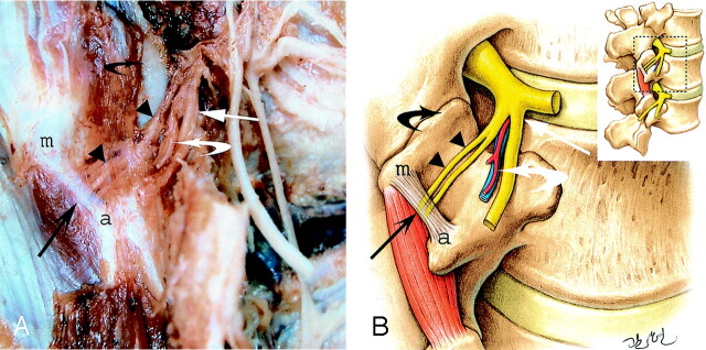
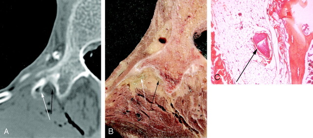
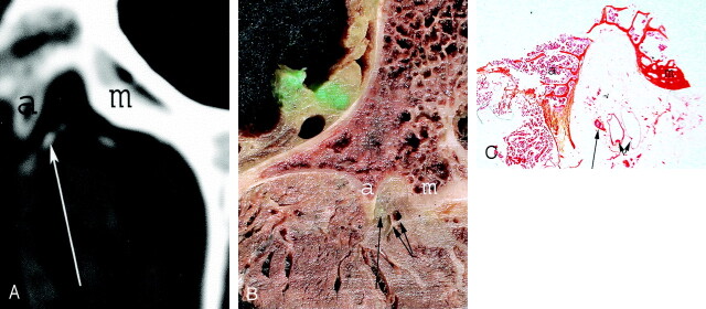
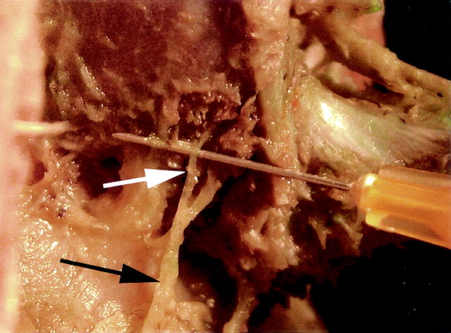
Methods: Lumbar spines of two cadavers were first dissected to assess the route of the L1-L5 posterior ramus. Thirty lumbar spinal nerves of three cadavers were injected in the epineural space with a mixture of iodine contrast and stain to perform a correlation between anatomic gross sections and CT sections in the axial plane. A histologic study was also performed to ensure the neurologic nature of the structure identified.
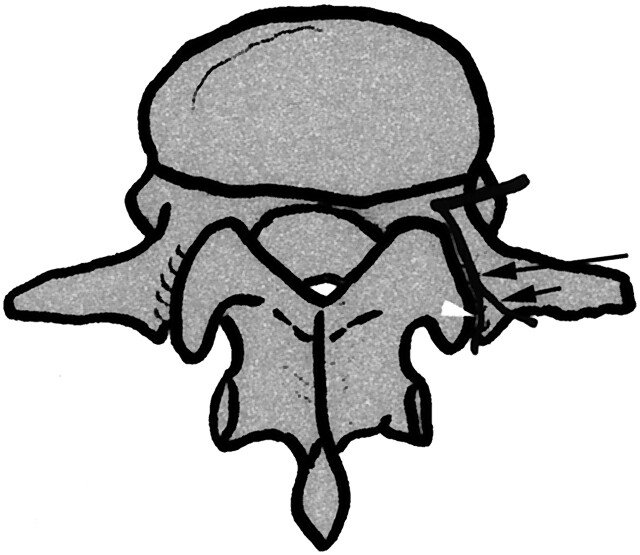
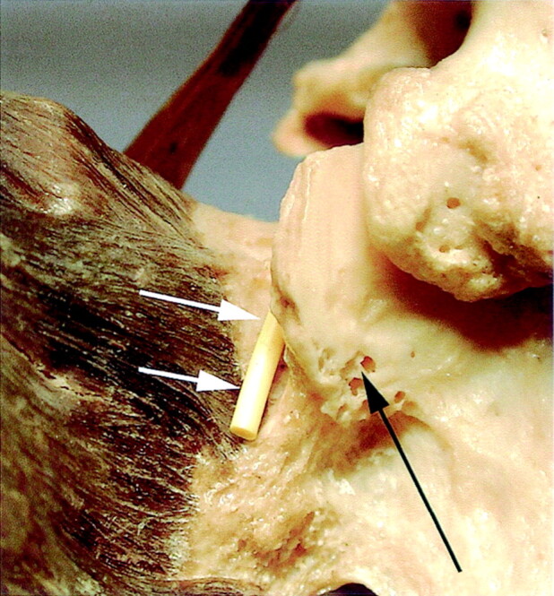
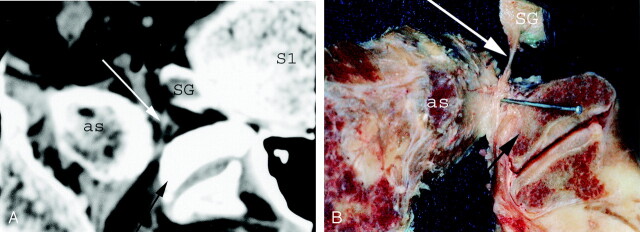
Results: The fibroosseous canal located between the mamillary and the accessory processes was a constant pathway for the medial branch of the L1-L4 posterior ramus. This former was always located closer to the accessory process. The L5 posterior ramus and its divisions could also be identified into a groove bounded laterally by the ala of the sacrum and medially by the base of the superior articular process of S1.
Conclusion: The accessory process and the groove bounded laterally by the ala of the sacrum and medially by the base of the superior articular process of S1 can be easily depicted on CT images and may allow a precise and selective infiltration of the medial branch of the posterior lumbar ramus.
Figures
Comment in
-
In vivo neuroanatomy and objectivity.AJNR Am J Neuroradiol. 2005 Apr;26(4):692. AJNR Am J Neuroradiol. 2005. PMID: 15814905 Free PMC article. No abstract available.
Similar articles
-
The anatomy of the so-called "articular nerves" and their relationship to facet denervation in the treatment of low-back pain.J Neurosurg. 1979 Aug;51(2):172-7. doi: 10.3171/jns.1979.51.2.0172. J Neurosurg. 1979. PMID: 156249
-
Proximal lumbar spinal nerves in axial MR imaging, CT, and anatomic sections.Radiology. 1992 Apr;183(1):239-41. doi: 10.1148/radiology.183.1.1549679. Radiology. 1992. PMID: 1549679
-
Extraforaminal entrapment of the fifth lumbar spinal nerve by osteophytes of the lumbosacral spine: anatomic study and a report of four cases.Spine (Phila Pa 1976). 2002 Mar 15;27(6):E169-73. doi: 10.1097/00007632-200203150-00020. Spine (Phila Pa 1976). 2002. PMID: 11884922
-
A new simplified sonographic approach for pararadicular injections in the lumbar spine: a CT-controlled cadaver study.AJNR Am J Neuroradiol. 2011 May;32(5):828-31. doi: 10.3174/ajnr.A2389. Epub 2011 Feb 24. AJNR Am J Neuroradiol. 2011. PMID: 21349957 Free PMC article.
-
Anatomy of the extradural compartments of the lumbar spinal canal. Peridural membrane and circumneural sheath.Radiol Clin North Am. 2000 Nov;38(6):1177-206. doi: 10.1016/s0033-8389(08)70003-4. Radiol Clin North Am. 2000. PMID: 11131629 Review.
Cited by
-
Comparison of the Efficacy of Different Radiofrequency Techniques for the Treatment of Lumbar Facet Joint Pain: Combined with Anatomy.Curr Pain Headache Rep. 2024 Jul;28(7):699-708. doi: 10.1007/s11916-024-01241-7. Epub 2024 Mar 25. Curr Pain Headache Rep. 2024. PMID: 38526650 Review.
-
Clinical Anatomy and Measurement of the Medial Branch of the Spinal Dorsal Ramus.Medicine (Baltimore). 2015 Dec;94(52):e2367. doi: 10.1097/MD.0000000000002367. Medicine (Baltimore). 2015. PMID: 26717379 Free PMC article.
-
All about pain pharmacology: what pain physicians should know.Korean J Pain. 2020 Apr 1;33(2):108-120. doi: 10.3344/kjp.2020.33.2.108. Korean J Pain. 2020. PMID: 32235011 Free PMC article. Review.
References
-
- Ghormley R. Low back pain with special reference to the articular facet, with presentation of an operative procedure. JAMA 1933;101:1773–1777
-
- Schellinger D, Werner L, Ragsdale B, Patronas NJ. Facet joint disorder and their role in production of back pain and sciatica. Radiographics 1987;7:923–944 - PubMed
-
- Helbig T, Casey K. Lee. The lumbar facet syndrome. Spine 1988;13:61–64 - PubMed
-
- Fukui S, Ohseto K, Shiotani M, et al. Distribution of referred pain from the lumbar zygapophyseal joints and dorsal rami. Clin J Pain 1997;13:303–307 - PubMed
-
- Lazorthes G. La branche postérieure des nerfs rachidiens: l’innervation des articulations interapophysaires vertébrales [in French]. In: Compte-rendu de l’Association des Anatomistes. 43ème reunion. Lisbonne:000;1966. :488–494
MeSH terms
LinkOut - more resources
Full Text Sources
Medical