. 2005 Apr;26(4):711-8.

Preangiographic evaluation of spinal dural arteriovenous fistulas with elliptic centric contrast-enhanced MR Angiography and effect on radiation dose and volume of iodinated contrast material

Affiliations

Preangiographic evaluation of spinal dural arteriovenous fistulas with elliptic centric contrast-enhanced MR Angiography and effect on radiation dose and volume of iodinated contrast material

Patrick H Luetmer et al. AJNR Am J Neuroradiol. 2005 Apr.

Abstract

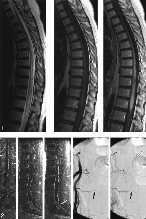
Background and purpose: The detection and localization of spinal dural arteriovenous fistulas (AVFs) remain diagnostic challenges. This study tested the hypothesis that elliptic centric contrast-enhanced MR angiography (MRA) can be used to detect spinal dural AVFs, predict the level of fistulas, and reduce the radiation dose and volume of iodinated contrast material associated with conventional angiography.

Methods: We examined 31 patients who presented with suspected spinal dural AVF between December 2000 and March 2004. All patients underwent MRA and conventional angiography. The effect of MRA on subsequent conventional angiography was assessed by analyzing total fluoroscopy time and volume of iodinated contrast material used.

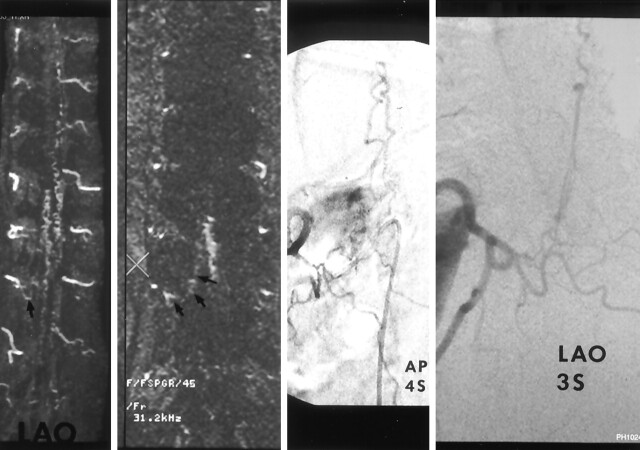
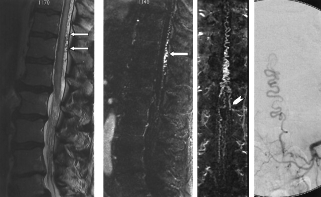
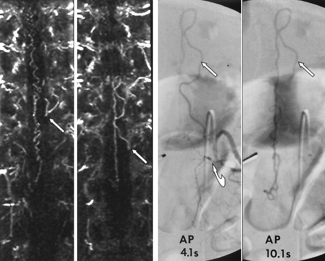
Results: At angiography, spinal dural AVFs were diagnosed in 22 of 31 patients, and MRA depicted an AVF in 20 of the 22 patients. MRA findings correctly predicted a negative angiogram in seven of nine cases. Of the 20 true-positive MRA results, the level of the fistula was included in the imaging volume in 14. In 13 of these 14 cases, MRA results correctly predicted the side and the level of the fistula to within one vertebral level. Fluoroscopy time and the volume of contrast agent was reduced by more than 50% in the 13 patients with a spinal dural AVF in whom MRA prospectively indicated the correct level.

Conclusion: Contrast-enhanced MRA can be used to detect spinal dural AVFs, predict the level of fistulas, and substantially reduce the radiation dose and volume of contrast agent associated with catheter spinal angiography.


Figures

F<sc>ig</sc> 1.
Fig 1.
Images in a 53-year-old woman with a 3-year history of progressive, burning pain; numbness; and subsequent weakness in her lower extremity. She had bladder incontinence in the last year. Left, Sagittal fast spin-echo image shows increased T2 signal intensity and mild swelling involving the lower cord and conus. Flow voids are present over the dorsal surface of the cord. Middle and right, Sagittal T1-weighted images without (middle) and with (right) gadolinium enhancement show patchy enhancement of the lower cord and conus. It is difficult to appreciate the prominent coronal venous plexus on the enhanced image.
F<sc>ig</sc> 2.
Fig 2.
Sagittal MRA performed with a bolus of gadolinium-based contrast agent. Thin, 6-mm coronal (far left) and sagittal (middle left and middle) MIP images obtained at 2-mm increments through the spinal canal show a prominent, tortuous medullary vein extending from the level of the lower body of S1 (white arrow) to the conus and a dilated coronal venous plexus. Catheter, right internal iliac angiograms (middle right and right) show a spinal dural AVF at S4 on the right (black arrows). Draining medullary vein is relatively straight until it begins to meander in the subarachnoid space at the level of the lower body of S1 (white arrow).
F<sc>ig</sc> 3.
Fig 3.
Images in a 72-year-old man with a 2-year history of urinary incontinence with progressive perianal and lower-extremity numbness and loss of lower-extremity control over the last year leading to use of a wheelchair in the last 2 months. Far left, MIP image in the left anterior oblique projection shows prominent, tortuous vessels on the dorsal and ventral surfaces of the cord and a prominent vessel extending from the right T11 foramen (arrow). Middle left, Source image confirms a prominent vessel (arrows) extending from below the right T11 pedicle (X) to prominent vessels on the surface of the cord. Middle right and right, Catheter angiograms in anteroposterior and left anterior oblique views at T11 on the right confirms a spinal dural AVF inferior to the right T11 pedicle draining to a dilated coronal venous plexus.
F<sc>ig</sc> 4.
Fig 4.
Images in a 64-year-old man with a 13-month history of progressive lower trunk and lower-extremity numbness and mild bowel and bladder urgency. Far left, Sagittal T2-weighted fast spin-echo image shows increased T2 signal intensity and mild swelling involving the lower spinal cord and conus, with prominent flow voids over the dorsal surface of the cord (arrows). Middle left and middle right, MRA performed with a bolus of gadolinium-based contrast agent. Thin, 6-mm sagittal (middle left) and coronal (middle right) MIP images at 2 mm increments through the spinal canal confirm a dilated coronal venous plexus along the dorsal surface of the cord (arrow) and show a prominent medullary vein extending from the left L1 foramen (arrowhead). Far right, Catheter angiogram confirms a spinal dural AVF at L1 on the left side.
F<sc>ig</sc> 5.
Fig 5.
Images in a 62-year-old man with a 19-month history of progressive lower-extremity motor and sensory dysfunction and neurogenic bladder diagnosed elsewhere, with transverse myelitis. Far left and middle left, MRA performed with a bolus of gadolinium-based contrast agent and thin, 6-mm coronal MIP images obtained at 2-mm increments through the spinal canal. Far left, Image shows a prominent medullary vein at the level of the left T11 foramen (arrow). Middle left, Image shows that the medullary vein is continuous with a prominent medullary vein emanating from the left T12 foramen (arrow). Far right and middle right, Limited catheter, 4- and 10-second spinal angiograms confirm a spinal dural AVF located under the left T12 pedicle (curved arrow). Left T12 medullary vein loops back toward the left T11 foramen (straight arrow). The 10-second delayed image shows opacification of the dilated coronal venous plexus. MRA allowed us to limit angiography to the bilateral T11-L1 segmental arteries and thus limit the cost, radiation exposure, and dose of contrast agent (to 57 mL).

Similar articles

Cited by

References

    1. Saraf-Lavi E, Bowen BC, Quencer RM, et al. Detection of spinal dural arteriovenous fistulae with MR imaging and contrast-enhanced MR angiography: sensitivity, specificity, and prediction of vertebral level. AJNR Am J Neuroradiol 2002;23:858–867 - PMC - PubMed
    1. Farb RI, Kim JK, Willinsky RA, et al. Spinal dural arteriovenous fistula localization with a technique of first-pass gadolinium-enhanced MR angiography: initial experience. Radiology 2002;222:843–850 - PubMed
    1. Anson JA, Spetzler RF. Classification of spinal arteriovenous malformations and implications for treatment. BNI Qtr 1992;8:2–8
    1. Gilbertson JR, Miller GM, Goldman MS, Marsh WR. Spinal dural arteriovenous fistulas: MR and myelographic findings. AJNR Am J Neuroradiol 1995;16:2049–2057 - PMC - PubMed
    1. Grote EH, Bien S. Arteriovenous malformations of the spinal cord. In: Youmans JR, ed. Neurological Surgery. 4th ed. Philadelphia: W.B. Saunders;1996. :1511–1530

Publication types

LinkOut - more resources