

Dilated perivascular spaces: hallmarks of mild traumatic brain injury
- PMID: 15814911
- PMCID: PMC7977096
Dilated perivascular spaces: hallmarks of mild traumatic brain injury
Erratum in
- AJNR Am J Neuroradiol. 2007 Feb;28(2):199
Abstract
Background and purpose: Recent animal and human studies have shown an increased frequency of enlarged, high-convexity Virchow-Robin spaces (VRS) in several neurologic diseases, suggesting their role as neuroradiologic markers of inflammatory changes. The aim of this study was to determine the prevalence of high-convexity dilated VRS in mild traumatic brain injury (TBI).
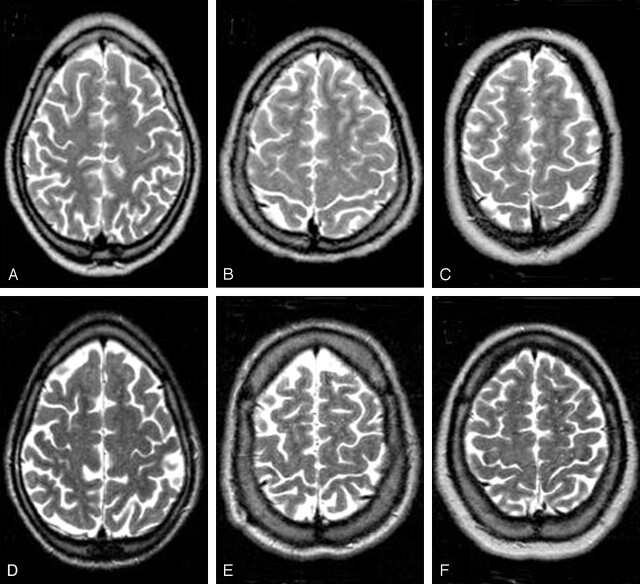
Methods: T2-weighted, T1-weighted, fluid-attenuated inversion recovery, and T2*-weighted gradient-echo brain MR images were acquired in 24 patients with TBI (10 women, 14 men; mean age, 33.6; range, 18.1-50.8 years) and 17 age- and sex-matched healthy control subjects (nine women, eight men; mean age, 32.8; range, 18.4-47.8 years). The mean interval after TBI was 3.6 days (range, 1-9 days) in 15 patients and 3.7 years (range, 0.6-13.4 years) in nine patients. Axial T2-weighted images were used to identify dilated VRS and to measure CSF volume; T1-weighted images were used to measure brain volume. Dilated VRS were identified as punctuate areas with CSF-like signal intensity in the high-convexity white matter.
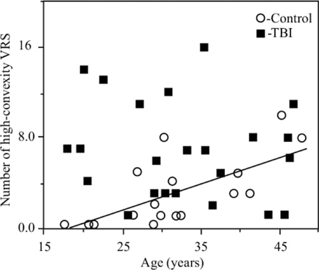
Results: Mean (+/- standard deviation) number of VRS was significantly higher in patients (7.1 +/- 4.6) than in controls (3.0 +/- 3.0, P = 0.002) [corrected] In controls, VRS were associated with age (R = 0.69, P < .001) whereas in patients, they neither correlated with brain and CSF volumes nor with age and the elapsed time from injury.
Conclusion: Our results suggest that the increased number of dilated VRS is a radiologic marker of mild head injury that is readily detectable on T2-weighted images. Because their number does not vary with time from injury, VRS probably reflect early and permanent brain changes.
Figures
References
Publication types
MeSH terms
Grants and funding
LinkOut - more resources
Full Text Sources