

MR cerebral blood volume maps correlated with vascular endothelial growth factor expression and tumor grade in nonenhancing gliomas
- PMID: 15814920
- PMCID: PMC7977110
MR cerebral blood volume maps correlated with vascular endothelial growth factor expression and tumor grade in nonenhancing gliomas
Abstract
Background and purpose: Relative cerebral blood volume (rCBV) measurements derived from perfusion-weighted imaging (PWI) may be useful to evaluate angiogenesis and preoperatively estimate the grade of a glioma. We hypothesized that rCBV is correlated with vascular endothelial growth factor (VEGF) expression as marker of the angiogenic stimulus in presumed supratentorial low-grade gliomas (LGGs).
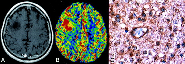
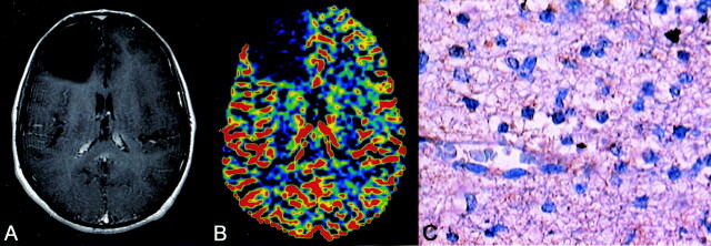
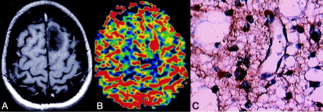
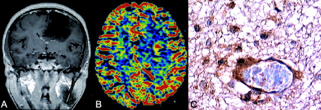
Methods: From February 2001 to February 2004, we examined 20 adults (16 men, four women; mean age 36 years; range, 23-60 years) with suspected (nonenhancing) supratentorial LGG on conventional MR imaging. Preoperative MR imaging used a dynamic first-pass gadolinium-enhanced, spin-echo echo-planar PWI. In heterogeneous tumors, we performed stereotactic biopsy in the high-perfusion areas before surgical resection. Semiquantitative grading of VEGF immunoreactivity was applied.
Results: Nine patients had diffuse astrocytomas (World Health Organization grade II), and 11 had other LGG and anaplastic gliomas. In patients with heterogeneous tumors on PWI, the high-rCBV focus had areas of oligodendroglioma or anaplastic astrocytoma on stereotactic biopsy, whereas the surgical specimens were predominantly astrocytomas. Anaplastic gliomas had high rCBV ratios and positive VEGF immunoreactivity. Diffuse astrocytomas had negative VEGF expression and mean rCBV values significantly lower than those of the other two groups. Three diffuse astrocytomas had positive VEGF immunoreactivity and high rCBV values.
Conclusion: Our results confirmed the correlation among rCBV measurements, VEGF expression, and histopathologic grade in nonenhancing gliomas. PWI may add useful data to the preoperative assessment of nonenhancing gliomas. Its contribution in predicting tumor behavior and patient prognosis remains to be determined.
Figures
References
-
- Kleihues P, Cayenee W. Pathology and Genetics of Tumors of the Nervous System. Lyon: International Agency for Research on Cancer;2000
-
- Wenz F, Rempp K, Hess T, et al. Effect of radiation on blood volume in low-grade astrocytomas and normal brain tissue: quantification with dynamic susceptibility contrast MR imaging. AJR Am J Roentgenol 1996;166:187–193 - PubMed
-
- Tomoi M, Maeda M, Yoshida M, et al. Assessment of radiotherapeutic effect on brain tumors by dynamic susceptibility contrast MR imaging: a preliminary report. Radiat Med 1999;17:195–199 - PubMed
-
- Pardo FS, Aronen HJ, Kennedy D, et al. Functional cerebral imaging in the evaluation and radiotherapeutic treatment planning of patients with malignant glioma. Int J Radiat Oncol Biol Phys 1994;30:663–669 - PubMed
MeSH terms
Substances
LinkOut - more resources
Full Text Sources
Other Literature Sources
Medical