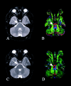
Diffusion tensor imaging of the corticospinal tract before and after mass resection as correlated with clinical motor findings: preliminary data
- PMID: 15814922
- PMCID: PMC7977087
Diffusion tensor imaging of the corticospinal tract before and after mass resection as correlated with clinical motor findings: preliminary data
Abstract
Background and purpose: The role of diffusion tensor imaging (DTI) in neurosurgical planning and follow-up is currently being defined and needs clinical validation. To that end, we sought correlations between preoperative and postoperative DTI and clinical motor deficits in patients with space-occupying lesions involving the corticospinal tract (CST).
Methods: DTI findings in four patients with masses near the CST and not involving motor cortex were retrospectively reviewed and compared with contralateral motor strength. CST involvement was determined from anisotropy and eigenvector directional color maps. The CST was considered involved if it was substantially deviated or had decreased anisotropy. Interpretations of the DTIs were blinded to assessments of motor strength, and vice versa.
Results: Of the four patients with potential CST involvement before surgery, DTI confirmed CST involvement in three, all of whom had preoperative motor deficits. The patient without CST involvement on DTI had no motor deficit. After surgery, DTI showed CST preservation and normalization of the position and/or anisotropy in two of the three patients with preoperative deficits, and both of those patients had improvement in motor strength. The other patient with preoperative deficits had evidence of wallerian degeneration on DTI and had only equivocal clinical improvement.
Conclusion: Preoperative CST involvement, as determined on DTI, was predictive of the presence or absence of motor deficits, and postoperative CST normalization on DTI was predictive of clinical improvement. Further study is warranted to define the role of DTI in planning tumor resections and predicting postoperative motor function.
Figures


References
-
- Tummala RP, Chu RM, Liu H, Truwit CL, Hall WA. Application of diffusion tensor imaging to magnetic-resonance-guided brain tumor resection. Pediatr Neurosurg 2003;39:39–43 - PubMed
-
- Field AS, Alexander AL, Wu YC, Hasan KM, Witwer B, Badie B. Diffusion tensor eigenvector directional color imaging patterns in the evaluation of cerebral white matter tracts altered by tumor. J Magn Reson Imaging 2004;20:555–562 - PubMed
-
- Mori S, Frederiksen K, van Zijl PCM, et al. Brain white matter anatomy of tumor patients evaluated with diffusion tensor imaging. Ann Neurol 2002;51:377–380 - PubMed
-
- Clark CA, Barrick TR, Murphy MM, Bell BA. White matter fiber tracking in patients with space-occupying lesions of the brain: a new technique for neurosurgical planning? Neuroimage 2003;20:1601–1608 - PubMed
Publication types
MeSH terms
Grants and funding
LinkOut - more resources
Full Text Sources
Medical
