

Mechanisms of bihemispheric brain infarctions in the anterior circulation on diffusion-weighted images
- PMID: 15814925
- PMCID: PMC7977111
Mechanisms of bihemispheric brain infarctions in the anterior circulation on diffusion-weighted images
Abstract
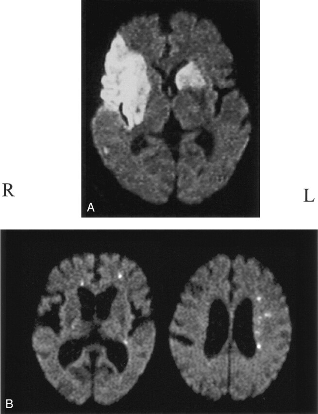
Background and purpose: Multiple acute brain infarctions in both cerebral hemispheres usually suggest an embolic mechanism, particularly one of aortic or cardiac origin. The purpose of this study was to clarify the etiologic mechanisms and topographic features of bihemispheric infarctions depicted on diffusion-weighted imaging (DWI).
Methods: Among 411 consecutive patients with ischemic stroke who underwent MR imaging in the acute phase, DWI showed bilateral infarctions in 19 (4.6%). In these patients, we analyzed the presence of carotid, aortic or cardiac embolic sources by using ultrasonography, cerebral angiography, and/or transesophageal echocardiography and evaluated the size and topographic distribution of the lesions. We assessed intracranial cross-flow through the anterior communicating artery, mainly on the basis of the anatomic information obtained from angiography or MR angiography.
Results: Bilateral lesions were derived from cardiac and/or aortic embolic sources in 16 (84%) of 19 patients and appeared to originate from unilateral carotid diseases in three (16%). In nine (82%) of 11 patients with cardiac embolic sources, at least one large territorial or subcortical lesion was found in either hemisphere, whereas in all eight patients without a cardiac embolic source, the lesions were small and disseminated bilaterally.
Conclusion: Unilateral carotid lesions can cause bihemispheric infarctions through cross-flow in the anterior communicating artery. On DWI, small bihemispheric, disseminated lesions strongly suggest an artery-to-artery embolism. In such cases, aortic and carotid lesions should be assessed as potential embolic sources.
Figures

Similar articles
-
Acute small subcortical infarctions on diffusion weighted MRI: clinical presentation and aetiology.J Neurol Neurosurg Psychiatry. 2005 Nov;76(11):1520-4. doi: 10.1136/jnnp.2005.063594. J Neurol Neurosurg Psychiatry. 2005. PMID: 16227543 Free PMC article.
-
[Multiple small subcortical infarction required to distinguish from lacunar infarction: evaluation by use of diffusion-weighted imaging].No To Shinkei. 2003 Dec;55(12):1041-6. No To Shinkei. 2003. PMID: 14870574 Japanese.
-
Bihemispheric subcortical infarcts in the middle cerebral artery territory.Clin Med Insights Case Rep. 2011;4:25-8. doi: 10.4137/CCRep.S7121. Epub 2011 Jun 1. Clin Med Insights Case Rep. 2011. PMID: 22084609 Free PMC article.
-
Differences in diffusion-weighted image and transesophageal echocardiographical findings in cardiogenic, paradoxical and aortogenic brain embolism.Cerebrovasc Dis. 2011;32(2):148-54. doi: 10.1159/000328652. Epub 2011 Jul 20. Cerebrovasc Dis. 2011. PMID: 21778712
-
Diffusion-weighted imaging-documented bilateral small embolic stroke involving multiple vascular territories may indicate occult cancer: A retrospective case series and a brief review of the literature.Aging Med (Milton). 2020 Mar 25;3(1):53-59. doi: 10.1002/agm2.12105. eCollection 2020 Mar. Aging Med (Milton). 2020. PMID: 32232193 Free PMC article. Review.
Cited by
-
Significance of Multiple Acute Ischemic Lesions on Initial Diffusion-weighted Imaging in Stroke Patients and Relation of Toast Classification.Ann Indian Acad Neurol. 2018 Jul-Sep;21(3):197-202. doi: 10.4103/aian.AIAN_487_17. Ann Indian Acad Neurol. 2018. PMID: 30258262 Free PMC article.
-
Disseminated Microinfarctions with Cerebral Microbleeds.J Stroke Cerebrovasc Dis. 2018 Jun;27(6):e95-e97. doi: 10.1016/j.jstrokecerebrovasdis.2017.12.032. Epub 2018 Feb 1. J Stroke Cerebrovasc Dis. 2018. PMID: 29395640 Free PMC article.
-
Beware of bihemispheric stroke after Omicron variant infection in the elderly.Brain Circ. 2023 Mar 24;9(1):52-54. doi: 10.4103/bc.bc_76_22. eCollection 2023 Jan-Mar. Brain Circ. 2023. PMID: 37151799 Free PMC article. No abstract available.
-
Bihemispheric ischemic strokes in patients with COVID-19.Brain Circ. 2022 Mar 21;8(1):10-16. doi: 10.4103/bc.bc_65_21. eCollection 2022 Jan-Mar. Brain Circ. 2022. PMID: 35372732 Free PMC article.
-
Mechanism of multiple infarcts in multiple cerebral circulations on diffusion-weighted imaging.J Neurol. 2007 Jul;254(7):924-30. doi: 10.1007/s00415-006-0397-3. Epub 2007 Apr 2. J Neurol. 2007. PMID: 17401747
References
-
- Altieri M, Metz RJ, Muller C, Maeder P, Meuli R, Bogousslavsky J. Multiple brain infarcts: Clinical and neuroimaging patterns using diffusion-weighted magnetic resonance. Eur Neurol 1999;42:76–82 - PubMed
-
- Baird AE, Lovblad KO, Schlaug G, Edelman RR, Warach S. Multiple acute stroke syndrome: marker of embolic disease? Neurology 2000;54:674–678 - PubMed
-
- Bogousslavsky J, Bernasconi A, Kumral E. Acute multiple infarction involving the anterior circulation. Arch Neurol 1996;53:50–57 - PubMed
-
- Roh JK, Kang DW, Lee SH, Yoon BW, Chang KH. Significance of acute multiple brain infarction on diffusion-weighted imaging. Stroke 2000;31:688–694 - PubMed
-
- Siebler M, Sitzer M, Rose G, Bendfeldt D, Steinmetz H. Silent cerebral embolism caused by neurologically symptomatic high-grade carotid stenosis: event rates before and after carotid endarterectomy. Brain 1993;116:1005–1015 - PubMed
Publication types
MeSH terms
LinkOut - more resources
Full Text Sources