. 2005 Mar;84(2-3):341-54.
doi: 10.1016/j.ejcb.2004.12.019.

Identification of a stem cell candidate in the normal human prostate gland

Affiliations

Identification of a stem cell candidate in the normal human prostate gland

Monika Schmelz et al. Eur J Cell Biol. 2005 Mar.

Abstract

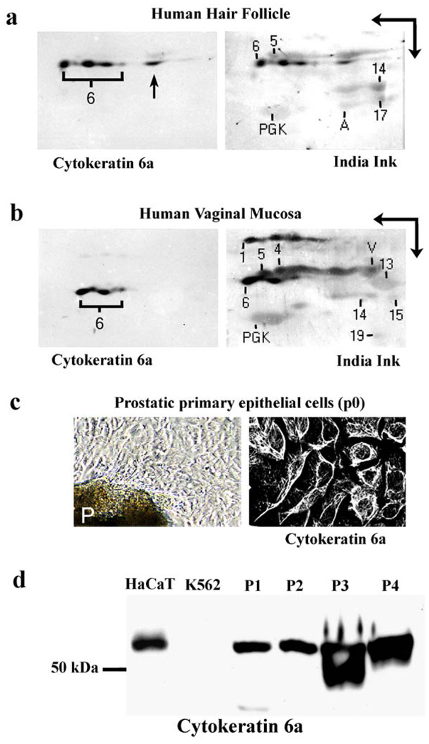
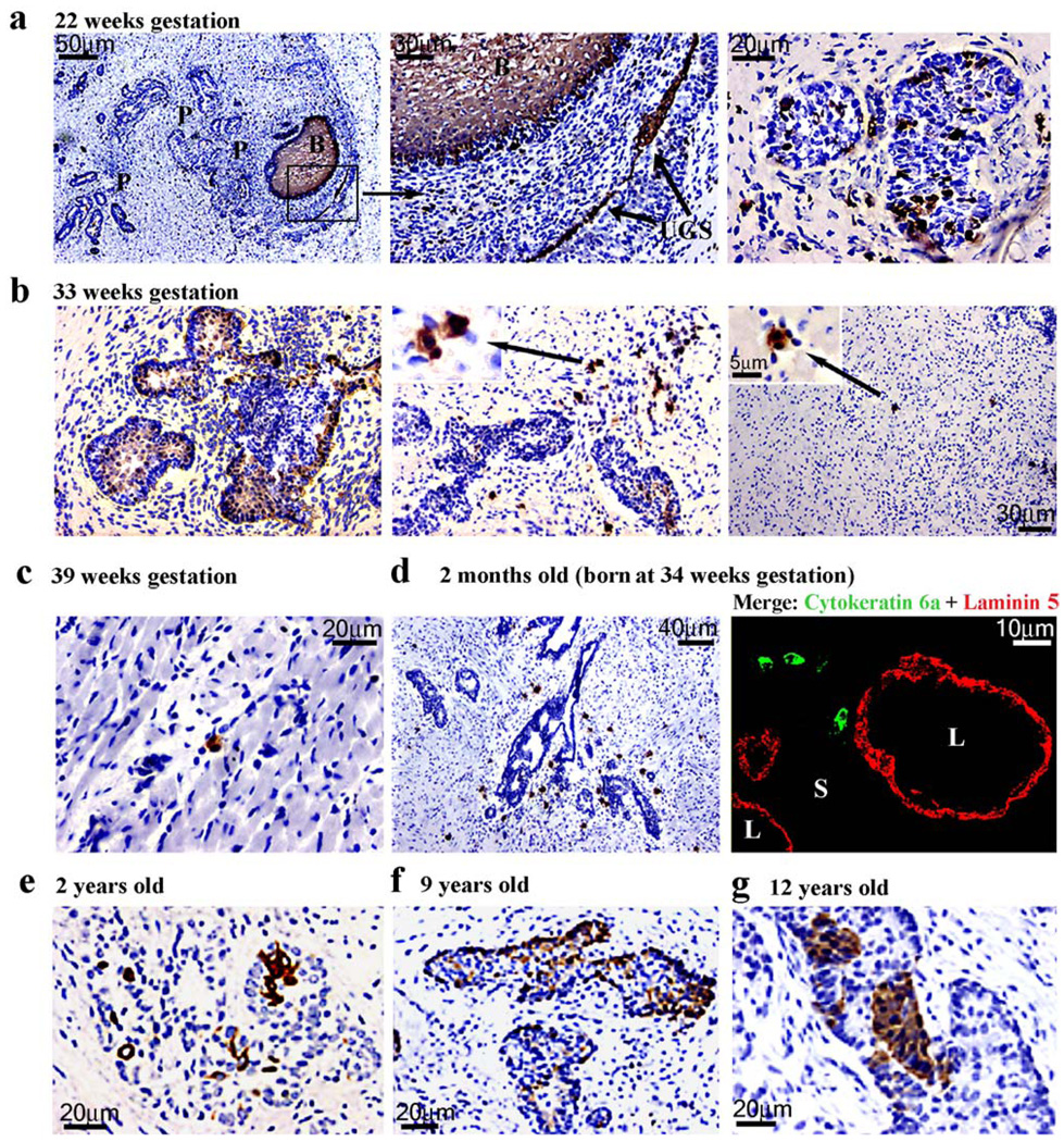
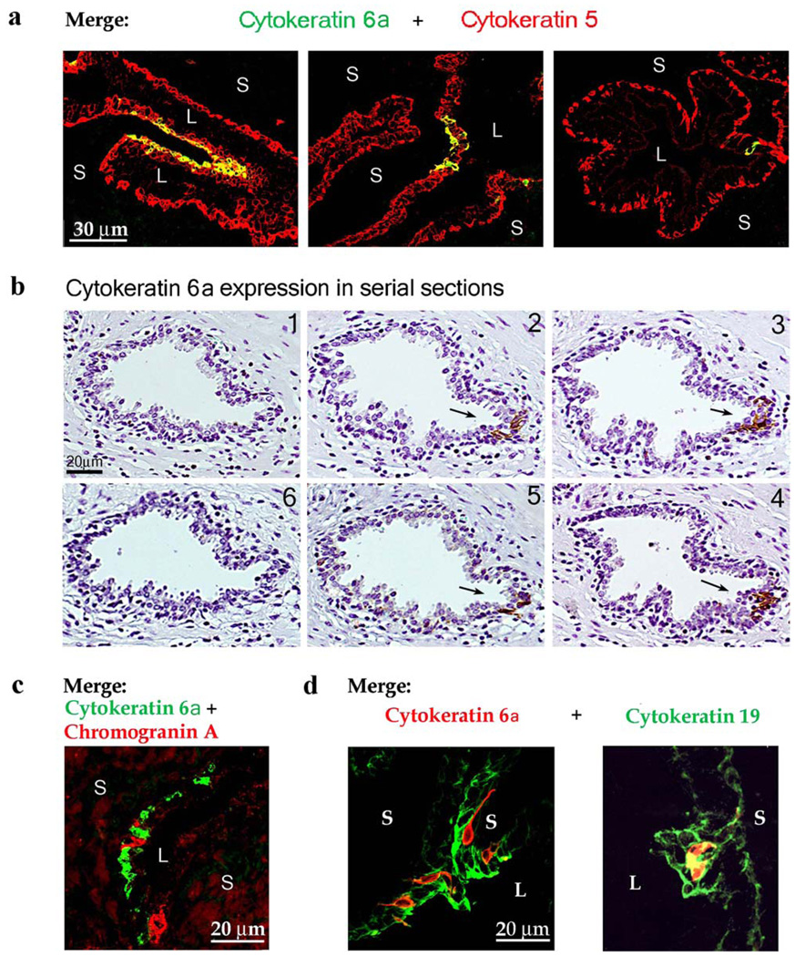
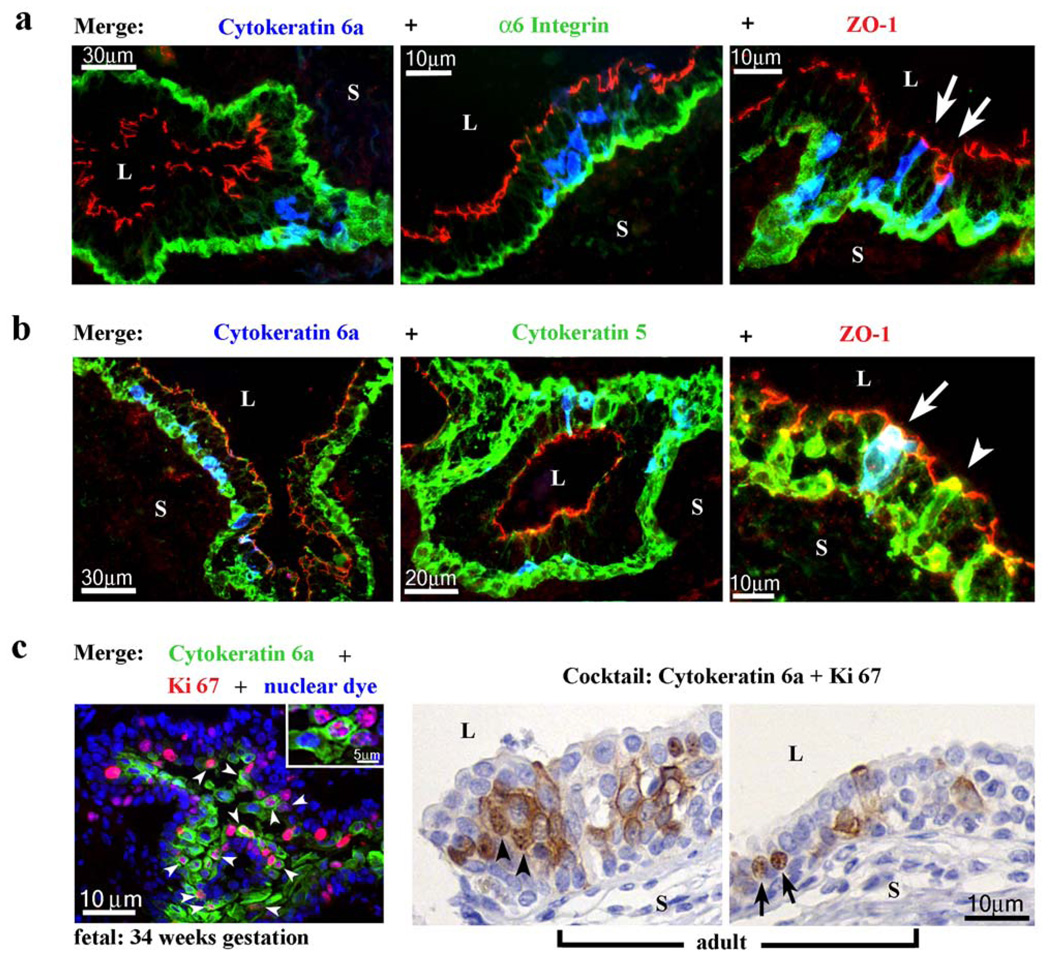
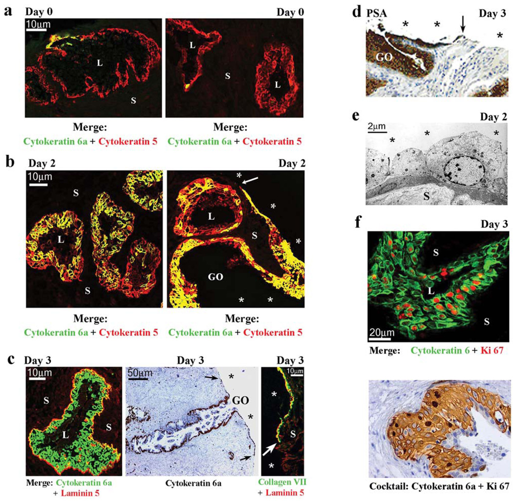
Stem cells of the human prostate gland have not yet been identified utilizing a structural biomarker. We have discovered a new prostatic epithelial cell phenotype-expressing cytokeratin 6a (Ck6a+ cells). The Ck6a+ cells are present within a specialized niche in the basal cell compartment in fetal, juvenile and adult prostate tissue, and within the stem cell-enriched urogenital sinus. In adult normal prostate tissue, the average abundance of Ck6a+ cells was 4.9%. With proliferative stimuli in the prostate organ culture model, in which the epithelial-stromal interaction was maintained, a remarkable increase of Ck6a expression was noticed to up to 64.9%. The difference in cytokeratin 6a expression between the normal adult prostate and the prostate organ culture model was statistically significant (p<0.0001). Within the prostate organ culture model the increase of cytokeratin 6a-expressing cells significantly correlated with increased proliferation index (r = 0.7616, p = 0.0467). The Ck6a+ cells were capable of differentiation as indicated by their expression of luminal cell markers such as ZO-1 and prostate specific antigen (PSA). Our data indicate that Ck6a+ cells represent a prostatic epithelial stem cell candidate possessing high potential for proliferation and differentiation. Since the development of benign prostatic hyperplasia and prostate carcinogenesis are disorders of proliferation and differentiation, the Ck6a+ cells may represent a major element in the development of these diseases.


Figures

Fig. 1
Fig. 1
Specificity of monoclonal antibody KA 12 for cytokeratin 6a. (a, b) Two-dimensional Western blotting (NEPHGE) of total protein extracts from the outer root sheath of human hair follicle (a) and from the human vaginal mucosa (b) with mAb KA 12. A = endogenous actin, V = endogenous vimentin, PGK = 3-phosphoglycerokinase added as protein marker, Numbers 1, 4–6, 13–15, 17 and 19 represent cytokeratins 1, 4–6,13–15,17,19 present in the total protein fraction. Double arrows in (a, b) denote the directions of the first dimension (horizontal arrow) and the second dimension (vertical arrow), respectively. Note that mAb KA12 reacted specifically with cytokeratin 6a including its isoelectric variants, and did not cross-react with any other cytokeratins present. The brackets indicate the isoelectric variants of cytokeratin 6a. Arrow in (a) denotes minor proportions of cytokeratins migrating in the first dimension as heterodimeric complexes. (c) The left panel shows an outgrowth of primary epithelial cells from a human prostatic tissue piece (P). The right panel shows cytokeratin-specific intermediate filament staining of mAb KA 12 in prostatic primary epithelial cells. Both images were taken at passage 0 (p0). (d) One-dimensional Western blot analysis showing the reaction of mAb KA 12 with cytokeratin 6a expressed in the primary epithelial cells (p0) obtained fromthe prostates of 4 patients (P1–P4). HaCaT cells were used as positive control, and K562 cells were used as negative control.
Fig. 2
Fig. 2
Cytokeratin 6a expression in the fetal (a–c) and juvenile (d–g) human prostate. (a) The prostatic ductal networks (P) at 22 weeks gestation (left image) are formed by solid epithelial outgrowths that emerge from the urogenital sinus (UGS) below the developing bladder epithelium(B = bladder). The framed area (middle image) and another distinct field with prostatic glands (right image) is shown at higher magnification. The UGS and the bladder epithelium abundantly expressed cytokeratin 6a. Some of the prostatic glandular epithelia expressed cytokeratin 6 in single cells or in cell clusters, whereas other glandular epithelial areas did not show any cytokeratin 6a expression. (b–c) At 33 (b) and 39 (c) weeks gestation, in addition to the cytokeratin 6a expression in glandular epithelium (left image), single or small groups of cytokeratin 6a-expressing cells were observed in the mesenchyme (middle and right image). Arrows indicate higher magnified fields (insets) of extraglandular cytokeratin 6a-expressing cells. These “extraglandular” cells were either in close proximity to glandular structures (middle image and inset) or distal from these (right image and inset). (d) Two-months old (born at 34 weeks gestation): The left panel shows mesenchymally located cytokeratin 6a cells in close proximity to a gland, which is cytokeratin 6a negative. The right image shows double-staining of cytokeratin 6a (green) with laminin 5 (red). (e–g) Shows cytokeratin 6a expression in the epithelium of juvenile prostatic glands. Cytokeratin 6a expression occurs in clusters or in single cells.
Fig. 3
Fig. 3
Cytokeratin 6a expression in normal human adult prostatic glands. (a) Three representative fields showing the distribution pattern of cytokeratin 6a-positive cells (green) by double-staining with basal cell-specific cytokeratin 5 (red), which outlines the basal compartment in normal glands. The fainter staining of luminal cells in the red channel is due to some cross-reaction with cytokeratins 8/18. Note that most cytokeratin 6a-expressing cells resided in the basal compartment. Co-localization of cytokeratin 6a with cytokeratin 5 appeared in yellow. (b) Immunohistochemical serial sections of one gland from the peripheral zone showing that cytokeratin 6a expression was restricted to so-called niches (arrows), a stem cell property. (c) Co-localization analysis of cytokeratin 6a (green) and chromogranin A (red) revealed that cytokeratin 6a-expressing cells are not identical with neuroendocrine cells. (d) Co-localization analysis of cytokeratin 6a (red) with cytokeratin 19 (green). Note that there is no co-localization in the one field, whereas the other field shows some co-localization appearing in yellow. S = stroma, L = glandular lumen.
Fig. 4
Fig. 4
Differentiation and proliferation capacity of cytokeratin 6a-positive cells in human normal prostate. (a) Three representative fields showing triple-staining of cytokeratin 6a (blue) with α6 integrin (green) and ZO-1 (red). Note that some cytokeratin 6aexpressing cells co-localized with α6 integrin in the basal compartment and thus appeared in turquoise (or lighter blue), while some other cytokeratin 6a cells also co-expressed ZO-1 (arrows). (b) Three representative fields showing triple-staining of cytokeratin 6a (blue) with cytokeratin 5 (green) and ZO-1 (red). Note that many cytokeratin 6a-expressing cells resided in the basal compartment by co-localization with cytokeratin 5 and thus appeared in turquoise (or lighter blue). A few cytokeratin 6a-expressing cells extended from the basal compartment to the lumen and co-expressed ZO-1 (red) and cytokeratin 5 (green). The overlay of green, red and blue appeared as white (arrow). Some cytokeratin 6a-negative but cytokeratin 5-positive cells also extended from the luminal compartment to the lumen and co-localized with ZO-1 appearing in yellow (arrowhead). (c) Triple-staining of cytokeratin 6a (green) with Ki 67 (red) and bisbenzimide H33258 (blue) in the fetal prostate (left image). Here, proliferative nuclei appeared in pink (insets). White arrowheads indicate proliferative cytokeratin 6a-positive cells. Two representative fields (middle and right image) with immunohistochemical staining using an anti-cytokeratin 6a (cytoskeletal) and anti-Ki 67 (nuclear) cocktail are shown for adult normal prostate. Black arrowheads indicate proliferating cells expressing cytokeratin 6a. Black arrows indicate proliferating cells that were cytokeratin 6a negative. S = troma, L = glandular lumen.
Fig. 5
Fig. 5
Amplification and proliferation potential of cytokeratin 6a-expressing cells. The prostate organ culture model revealed the amplification (a–c), differentiation (c–e) and proliferation potential (f) of Ck6a+ cells. (a) Prior to culturing (Day 0), the cytokeratin 6a expression pattern in the tissue sections is similar to that observed in vivo, as shown in two representative fields. Double-staining of cytokeratin 6a (green) with cytokeratin 5 (red) revealed a few clusters of Ck6a+ cells in the basal compartment of a few glands. (b) After Day 2 in culture, cytokeratin 6a expression was up-regulated in most glands as shown by double-staining with cytokeratin 5 (red) in two representative fields. Co-localization of these two antigens appeared as yellow. (c) After Day 3 in culture, most glands (left image) showed an abundant expression of cytokeratin 6a (green) as shown by double-staining with basement membrane specific laminin 5 (red). From cut open glands (GO), the Ck6a+ cells migrated to the surface and formed a new surface epithelium (*) as shown in the right image in (b) and by immunohistochemistry in the middle image in (c). Double-staining of laminin 5 (red) and collagen VII (green) showing that the new surface epithelium deposits these two proteins into a new extracellular matrix (right image in (c)). (d) shows PSA expression in the newly formed surface epithelium. (e) Electron micrograph of an area in the newly formed surface epithelium (*) showing differentiation into basal and luminal cells. (f) The up-regulation of cytokeratin 6a expression in this prostate organ model corresponded to the onset of proliferation as shown by double-staining of cytokeratin 6a (green) with Ki 67 (red) at Day 2, and by immunohistochemistry using a cocktail of anti-cytokeratin 6a (cytoskeletal) and anti-Ki 67 (nuclear) antibodies followed by visualization with DAB at Day 3. All white and black arrows point to the leading edge of the newly forming surface epithelium. All white and black stars indicate the free surface. S = stroma, L = glandular lumen, GO = gland cut open by coring of the prostatic tissue.

Similar articles

Cited by

References

    1. Achtstätter T, Hatzfeld M, Quinlan R, Parmelee D, Franke WW. Separation of cytokeratin polypeptides by gel electrophoretic and chromatographic techniques and their identification by immunoblotting. Methods Enzymol. 1986;134:355–371. - PubMed
    1. Barnett D, Huang H, Wu X, Laciak R, Shapir E, Bushman W. The human prostate expresses sonic hedgehog during fetal development. J. Urol. 2002;168:2206–2210. - PubMed
    1. Bierie B, Nozawa M, Renou JP, Shillingford JM, Morgan F, Oka T, Taketo MM, Cardiff RD, Miyoshi K, Wagner KU, Robinson GW, Hennighausen L. Activation of beta-catenin in prostate epithelium induces hyperplasias and squamous transdifferentiation. Oncogene. 2003;22:3875–3887. - PubMed
    1. Bjerknes M, Cheng H. Clonal analysis of mouse intestinal epithelial progenitors. Gastroenterology. 1999;116:7–14. - PubMed
    1. Bonkhoff H, Remberger K. Differentiation pathways and histogenetic aspects of normal and abnormal prostatic growth: a stem cell model. Prostate. 1996;28:98–106. - PubMed

Publication types