. 2005 May;115(1):42-54.
doi: 10.1111/j.1365-2567.2005.02127.x.

Epicutaneous immunization induces alphabeta T-cell receptor CD4 CD8 double-positive non-specific suppressor T cells that inhibit contact sensitivity via transforming growth factor-beta

Affiliations

Epicutaneous immunization induces alphabeta T-cell receptor CD4 CD8 double-positive non-specific suppressor T cells that inhibit contact sensitivity via transforming growth factor-beta

Marian Szczepanik et al. Immunology. 2005 May.

Abstract

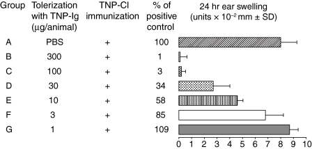
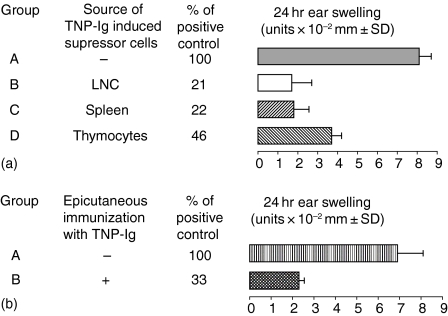
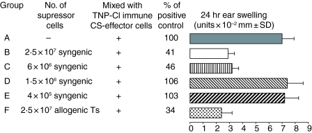
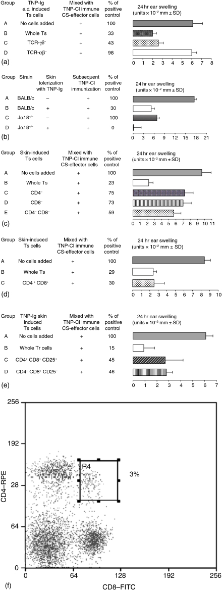
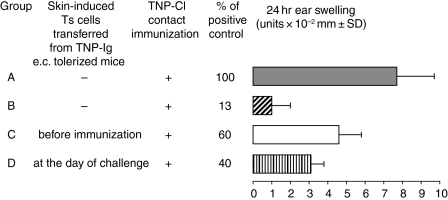
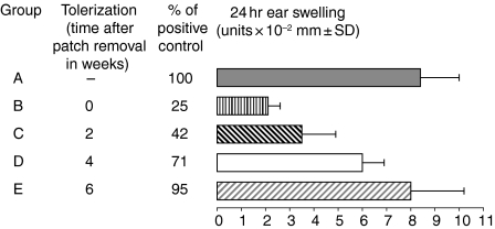
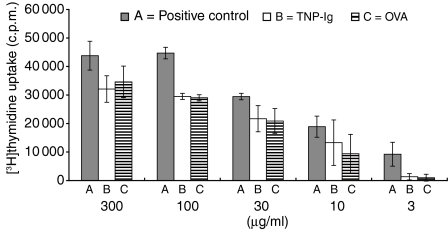
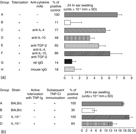
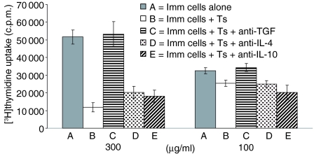
Since it was previously shown that protein antigens applied epicutaneously in mice induce allergic dermatitis mediated by production of T helper 2 (Th2) cytokines we postulated that this might induce suppression of Th1 immunity. Here we show that epicutaneous immunization of normal mice with a different protein antigen applied on the skin in the form of a patch induces a state of subsequent antigen-non-specific unresponsiveness caused by suppressor T cells (Ts) that inhibit sensitization and elicitation of effector T-cell responses. Suppression is transferable in vivo by alphabeta-T-cell receptor CD4(+) CD8(+) double positive lymphocytes harvested from lymphoid organs of skin patched animals and are not major histocompatibility complex-restricted nor antigen specific. Both CD25(+) and CD25(-) CD4(+) CD8(+) T cells are able to suppress adoptive transfer of Th1 effector cells mediating cutaneous contact sensitivity. In vivo treatment with monoclonal antibodies showed that the cytokines interleukin (IL)-4, IL-10 and transforming growth factor-beta (TGF-beta) are involved in the induction of the Ts cells. Additionally, using IL-10(-/-) mice we found that IL-10 is involved in skin induced tolerance. Further in vitro experiments showed that lymph node cells of skin tolerized mice non-specifically suppress [(3)H]thymidine incorporation by antigen-stimulated immune cells and this effect can be abolished by adding anti-TGF-beta, but not anti-IL-4 nor anti-IL-10 antibodies. These studies indicate the crucial role of TGF-beta in skin induced tolerance due to non-antigen-specific Ts cells and also show that IL-4, IL-10 and TGF-beta play an important role in the induction of epicutaneously induced Ts cell suppression.


Figures

Figure 1
Figure 1
Immunization (e.c) with protein antigen induces suppression of CS, which is dose dependent. CBA/J mice were e.c. immunized via patches with graded doses of TNP-Ig (300, 100, 30, 10, 3 or 1 µg/animal) (groups B–G) or exposed to PBS alone (group A). On day 7 patches were removed, mice were actively immunized with 5% TNP-Cl and then tested for CS. Results were expressed in units of 10−2 mm ± SD. Each experimental group consisted of five or six mice. Statistical significance: groups B, C, D and E versus group A; P < 0·001.
Figure 2
Figure 2
Skin-induced tolerance can be transferred into naïve recipients. (a) Skin induced tolerance can be transferred with cells of the immune system (‘transfer out’ protocol). Four day TNP-Cl immune cells (7 × 10⁷) were incubated for 30 min at 37° in medium alone (group A) or with 5 × 10⁷ peripheral LNC, spleen cells, or thymocytes isolated from mice that were tolerized with TNP-Ig (groups B–D). Then resultant cell mixtures were transferred into naive syngeneic recipients that were tested for CS. Each experimental group consisted of five to six mice. Statistical significance: groups B, C and D versus group A; P < 0·001.(b) Immunization of (e.c.) cell transfer recipients with protein antigen induces a suppressor environment that inhibits CS (transfer in protocol). To confirm data found in the cell mixing assay (‘transfer out’), 4 day TNP-Cl immune cells (7 × 10⁷) were transferred i.v. into syngeneic recipients that were tolerized by skin patching (group B) or into naïve mice (positive transfer) (group A). Then mice were challenged and tested for CS. Each experimental group consisted of five to six mice. Statistical significance: group B versus group A; P < 0·001.
Figure 3
Figure 3
A low number of suppressor cells is required to inhibit CS; 7 × 10⁷ 4 day TNP-Cl immune cells were incubated with decreasing numbers of syngeneic suppressor cells [CBA/J (H-2k)] (groups B–E) or with 2 × 10⁷ of allogenic Ts [BALB/c (H-2d)] or alone (group A) before cell transfer into naive recipients that were tested for CS. Statistical significance: groups B, C and F versus group A; P < 0·001.
Figure 4
Figure 4
Phenotype of Ts cells induced via e.c. immunization with protein antigen. (a) αβ-TCR+ lymphocytes are involved in skin induced tolerance. LN cells from mice e.c. immunized with TNP-Ig were treated with the following mAb: anti-TCR αβ, anti-TCR γδ (a); anti-CD4 or anti-CD8 (c) and RC or complement alone as a suppression control. Then 5 × 10⁷ suppressor cells treated with RC alone or cell equivalents of suppressor cells treated with appropriate mAb and RC, resultant cells were washed and incubated with 4 day TNP-Cl immune cells before transfer into syngeneic naive recipients that were tested for CS. Presented data show that skin induced suppressor cells belong to the population of TCR αβ+ (group C versus group B). Statistical significance: groups C and B versus group A; not significant; group D versus group A; not significant.(b) CD1d-dependent Vα14+ Jα18+ NKT cells are not required for skin induced tolerance. Additionally, experiments employing Jα18−/− mice e.c. immunized with TNP-Ig show that NKT cells are not involved in skin induced tolerance (group C versus D). Statistical significance: group C versus group D; P < 0·001. (c) Depletion of T cells that express CD4 and CD8 coreceptors abrogates the activity of skin induced suppressor cells. Presented data show that depletion of both CD4 and CD8 cells resulted in abrogation of suppressor activity induced via e.c. immunization (groups C and D versus group B). Additionally, reconstitution of CD4 depleted cells with the CD8 depleted cell population did not restore suppressor activity of skin induced Ts (group E versus group B). Statistical significance: group B versus group A; P < 0·001 and group D versus group C; not significant. (d) Epicutaneous immunization induces CD4+ CD8+ double-positive Ts cells.To determine if skin induced Ts cells coexpress CD4 and CD8, we incubated 2 × 10³ of sorted CD4+ CD8+ double-positive cells isolated from skin-tolerized mice with 4 day TNP-Cl immune cells before transfer into syngeneic naive recipients that were tested for CS. The data show that only 2 × 10³ CD4+ CD8+ double positive cells were able to efficiently suppress CS at the same level as 5 × 10⁷ unseparated regulatory cells (groups C and B versus group A). Statistical significance: groups C and B versus group A; P < 0·001. (e) Both CD25+ and CD25 CD4+ CD8+ cells from e.c.-immunized mice are able to inhibit adoptive transfer of CS. Presented data show that FACS-sorted CD4+ CD8+ cells that either CD25+ or CD25 can suppress adoptive transfer of CS (groups C and D versus group A). Statistical significance: group B versus group A; P < 0·001; group C versus group A; P < 0·02 and group D versus group A; P < 0·001. (f) Epicutaneous immunization with protein antigen causes increase of CD4+ CD8+ DP cells in the periphery. Spleen cells from mice epicutaneously immunized with TNP-Ig double-stained with FITC anti-CD8 and RPE anti-CD4 and analysed by flow cytometry. Lymphocytes were gated according to size and granularity on FSC versus SSC dotplot and the percentage of double positive CD4+ CD8+ cells within lymphocytes gate is presented. Results are representative of three independent experiments.
Figure 5
Figure 5
Transfer of regulatory cells both at the day of immunization or challenge can suppress CS. Regulatory cells isolated from TNP-Ig patched mice were transferred into syngeneic mice at the day of TNP-Cl immunization (day 0) or at the day of challenge (day 4) (groups C and D). There were two additional groups: positive control (mice actively immunized with TNP-Cl) (group A) and a group of mice that were patched with TNP-Ig and then actively sensitized with TNP-Cl (suppression control) (group B). Statistical significance: group B versus group A; P < 0·001; group C versus group A; P < 0·05 and group D versus group A; P < 0·01.
Figure 6
Figure 6
Skin induced suppression declines with time. CBA/J mice were e.c. immunized with 100 µg/animal of TNP-Ig or exposed to PBS alone (group A) as described previously. On day 7 patches were removed and then mice were actively immunized with 5% TNP-Cl immediately (group B), or 2, 4 or 6 weeks later (groups C-E correspondingly). Mice from all experimental groups were challenged and tested for CS. Statistical significance: groups B and C versus group A; P < 0·001; group D versus group A; P < 0·02.
Figure 7
Figure 7
The influence of e.c. tolerization on the in vitro proliferation of TNP-Cl immune lymph node cells. CBA/J mice were skin tolerized with TNP-Ig (group B), OVA (group C) or treated with PBS alone (group A). On day 7 patches were removed and mice from all groups were actively immunized with 5% TNP-Cl. Four days later axillary and inginual LN were collected and single-cell suspensions prepared and tested in proliferation assay. 3 × 10⁵ LNC per well were cultured in U-bottom 96-well microplates in triplicate in RPMI-1640 plus FCS, l-glutamine, HEPES and 2-mercaptoethanol. Threefold dilutions of TNP-Ig were added to the wells (consecutively 300, 100, 30, 10 and 3 µg/ml). After addition of 0·5 µCi/well of [3H]thymidyne, the cells were incubated for 18 h and then [3H]thymidyne incorporation was measured in a β scintillation counter. Results are expressed as mean ± SD. Statistical significance: 100 µg/ml, groups B and C versus group A; P < 0·05; 30 µg/ml groups B and C versus group A; P < 0·02. Differences in other groups are non-significant.
Figure 8
Figure 8
Inhibitory cytokines involved in the induction of skin induced suppressor T cells. (a) IL-4, IL-10 and TGF-β are required for induction of Ts cells via e.c. immuization. Mice were e.c. tolerized with TNP-Ig alone (group B) or TNP-Ig plus 100 µg of anticytokine mAb (anti-IL4, anti-IL-10, anti-TGF-β or the mixture of all three mAbs) (groups C-F, respectively). Then mice were actively immunized with 5% TNP-Cl and tested for CS. An additional group of mice were patched with PBS alone and then actively immunized with TNP-Cl (positive control) (group A). Additionally, in groups G and H mice were e.c. tolerized with TNP-Ig plus 100 µg of rat or mouse IgG, respectively, as an isotype control. Statistical significance: group B versus group A; P < 0·001, groups C and F versus group B; P < 0·001, group D versus group B; P < 0·05 and group E versus group B; P < 0·01. groups G and H versus group B; not significant. (b)IL-10 is involved in skin-induced tolerance. To confirm involvement of IL-10 in the induction of Ts cells via e.c. immunization with protein antigen we compared immune response in wild type BALB/c and IL-10-/— mice that were e.c. immunized with TNP-Ig (groups B and D) before active immunization with TNP-Cl. In groups A and C BALB/c and IL-10−/− mice were patched with PBS alone before TNP-Cl sensitization (positive controls). Statistical significance: group B versus group A; P < 0·001 and group D versus group B; P < 0·05.
Figure 9
Figure 9
Skin induced tolerance operates via αβ-TCR+ cells producing TGF-β. To determine the mechanism of skin induced tolerance, 6 × 10⁴ mitomycin C-treated LNC from tolerized mice (Ts) were cocultured with 3 × 10⁵ 4 day TNP-Cl immune cells (Imm. cells) in triplicates with or without following anti-cytokine mAbs: anti-IL4, anti-L-10 or anti-TGF-β. In the positive control immune cells were coultered without Ts cells. Conditions of culture and [3H]thymidyne were described in the legends to Fig. 7. Statistical significance: 300 µg/ml groups B, D and E versus group A; P < 0·05. Differences in other groups are non-significant.

References

    1. Weiner HL, Friedman A, Miller A, et al. Oral tolerance: Immunologic mechanisms and treatment of animal and human organ-specific autoimmune diseases by oral administration of autoantigens. Annu Rev Immunol. 1994;12:809–37. 10.1146/annurev.iy.12.040194.004113. - DOI - PubMed
    1. Xiao B-G, Link H. Mucosal tolerance: a two-edged sword to prevent and treat autoimmune diseases. Clin Immunol Immunopathol. 1997;85:119–28. 10.1006/clin.1997.4432. - DOI - PubMed
    1. Chen YJ, Inobe I, Kuchroo VK, Baron JL, Janeway CA, Weiner HL. Oral tolerance in myelin basic protein T-cell receptor trangenic mice. Suppression of autoimmune encephalomyelitis and dose-dependent induction of regulatory cells. Proc Natl Acad Sci USA. 1996;93:388–99. - PMC - PubMed
    1. Spergel JM, Mizoguchi E, Oettgen H, Bhan AK, Geha RS. Roles of Th1 and Th2 cytokines in a murine model of allergic dermatitis. J Clin Invest. 1999;103:1103–11. - PMC - PubMed
    1. Herrick CA, MacLeod H, Glusac E, Tigelaar RE, Bottomly K. Th2 responses induced by epicutaneous or inhalational protein exposure are differentially dependent on IL-4. J Clin Invest. 2000;105:765–75. - PMC - PubMed

Publication types

MeSH terms

Substances

LinkOut - more resources