. 2005 May;115(1):55-62.
doi: 10.1111/j.1365-2567.2005.02130.x.

The role of macrophages in the in vitro generation of extracellular DNA from apoptotic and necrotic cells

Affiliations

The role of macrophages in the in vitro generation of extracellular DNA from apoptotic and necrotic cells

Jin-Jung Choi et al. Immunology. 2005 May.

Abstract

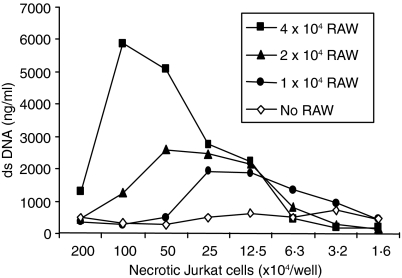
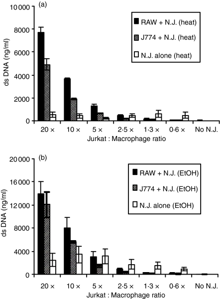
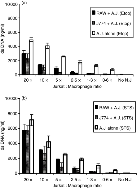
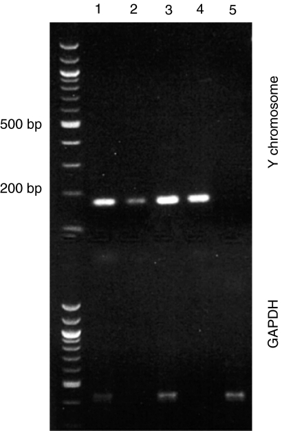
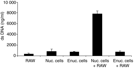
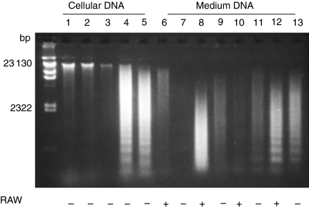
Cell death is a ubiquitous process that occurs by apoptosis or necrosis depending on the triggering event. While apoptotic and necrotic cells differ biochemically, both are cleared by macrophages for elimination. The process is very efficient, although DNA can appear in the blood in various clinical conditions associated with cell death. To define the role of macrophages in the generation of extracellular DNA, in vitro experiments were performed, assessing the release of DNA into the media of apoptotic or necrotic Jurkat cells cultured with RAW264.7 or J774 macrophage cell lines. DNA was measured by a fluorimetric assay using the dye PicoGreen. In these experiments, while necrotic cells alone did not release DNA, in the presence of macrophages, significant amounts of DNA appeared in the medium. This DNA contained sequences from the Jurkat cells and had reduced molecular weight in comparison to cellular DNA. Furthermore, coculture of macrophages with enucleated necrotic Jurkat cells did not release DNA, suggesting that the DNA came from the dead cell. In contrast, Jurkat cells made apoptotic by treatment with either staurosporine or etoposide spontaneously released DNA, while coculture with macrophages caused a decrease in the DNA released. With apoptotic cells, the DNA in the medium showed low molecular weight and laddering whether or not macrophages were present. Together, these results indicate that macrophages play an important role in the generation of extracellular DNA from dead and dying cells, with the effect dependent on how the cell died.


Figures

Figure 1
Figure 1
DNA release effects of macrophages cultured with necrotic Jurkat cells. Various numbers of RAW264.7 cells (1 × 10⁴, 2 × 10⁴, 4 × 10⁴/well) were cocultured with various numbers of heat-treated Jurkat cells (56°), and DNA levels in the media were determined by PicoGreen after 24 hr. The results presented are representative of three independent experiments.
Figure 2
Figure 2
DNA release from cocultures of macrophages with necrotic Jurkat cells. Following induction of necrosis with heat (56° for 30 min; a) or with 70% ethanol (EtOH; b), the amount of released DNA was measured in the media of RAW264.7 cells (4 × 10⁴/well) cocultured with the necrotic Jurkat cells at various doses for up to 48 hr using a fluorometric assay. Results (mean ± SD) were obtained from two experiments, each in triplicate.
Figure 3
Figure 3
DNA release from cocultures of macrophages with apoptotic Jurkat cells. Following induction of apoptosis with etoposide (Etop, 30 μg/ml; a) or staurosporine (STS, 5 μm; b), the amount of DNA was measured in the media of RAW264.7 cells (4 × 10⁴/well) cocultured with these apoptotic Jurkat cells at various doses up to 48 hr using a fluorometric assay. Results (mean ± SD) were obtained from two experiments, each in triplicate.
Figure 4
Figure 4
The origin of DNA released into the medium. The released DNA from various media as indicated was extracted and analysed on a 1% agarose gel. These DNAs were used as templates in polymerase chain reactions to amplify the Y chromosome and GAPDH gene. Lane 1, RAW264.7 with heated Jurkat cells; lane 2, heated Jurkat cells alone; lane 3, RAW264.7 with ethanol-treated Jurkat cells; lane 4, ethanol-treated Jurkat cells alone; lane 5, RAW264.7 cells alone.
Figure 5
Figure 5
The effect of enucleated cells on DNA release of macrophages. Enucleated (Enuc.) or nucleated (Nuc.) Jurkat cells were added to RAW264.7 cells (4 × 10⁴/well) with a ratio of 20 : 1, and DNA levels in the media were determined after 24 hr by fluorometric assay. Results (mean ± SD) were obtained from two experiments, each in triplicate.
Figure 6
Figure 6
The size of the released DNA. Cellular (lane 1–5) and extracellular (lanes 6–13) DNA from necrotic or apoptotic Jurkat cells were purified and analysed by gel electrophoresis with λ DNA digested with HindIII as a standard (left). Cellular DNA from untreated living (lane 1), heated (lane 2), ethanol-treated (lane 3), etoposide-treated (lane 4) and staurosporine-treated (lane 5) Jurkat cells are shown. Extracellular DNA was obtained from the media of RAW264.7 cells with heated Jurkat cells (lane 6); heated Jurkat cells alone (lane 7); RAW264.7 with ethanol-treated Jurkat cells (lane 8); ethanol-treated Jurkat cells alone (lane 9); RAW264.7 with etoposide-treated Jurkat cells (lane 10); etoposide-treated Jurkat cells alone (lane 11); RAW264.7 with staurosporine-treated Jurkat cells (lane 12); and staurosporine-treated Jurkat cells alone (lane 13). This experiment was repeated three times with similar results.

Similar articles

Cited by

References

    1. Majno G, Joris I. Apoptosis, oncosis, and necrosis: an overview of cell death. Am J Pathol. 1995;146:3–15. - PMC - PubMed
    1. Farber E. Programmed cell death: necrosis versus apoptosis. Modern Pathol. 1994;7:605–9. - PubMed
    1. Columbano A. Cell death: current difficulties in discriminating apoptosis from necrosis in the context of pathological processes in vivo. J Cell Biochem. 1995;58:181–90. - PubMed
    1. Martelli AM, Zweyer M, Ochs RL, et al. Nuclear apoptotic changes: an overview. J Cell Biochem. 2001;82:634–46. 10.1002/jcb.1186. - DOI - PubMed
    1. Ren Y, Savill J. Apoptosis: the importance of being eaten. Cell Death Differ. 1998;5:563–8. 10.1038/sj.cdd.4400407. - DOI - PubMed

Publication types

LinkOut - more resources