. 2005 Apr 8;6(1):31.
doi: 10.1186/1465-9921-6-31.

Deficiency in type 1 insulin-like growth factor receptor in mice protects against oxygen-induced lung injury

Affiliations

Deficiency in type 1 insulin-like growth factor receptor in mice protects against oxygen-induced lung injury

Karmene Ahamed et al. Respir Res. .

Abstract

Background: Cellular responses to aging and oxidative stress are regulated by type 1 insulin-like growth factor receptor (IGF-1R). Oxidant injury, which is implicated in the pathophysiology of a number of respiratory diseases, acutely upregulates IGF-1R expression in the lung. This led us to suspect that reduction of IGF-1R levels in lung tissue could prevent deleterious effects of oxygen exposure.

Methods: Since IGF-1R null mutant mice die at birth from respiratory failure, we generated compound heterozygous mice harboring a hypomorphic (Igf-1rneo) and a knockout (Igf-1r-) receptor allele. These IGF-1Rneo/- mice, strongly deficient in IGF-1R, were subjected to hyperoxia and analyzed for survival time, ventilatory control, pulmonary histopathology, morphometry, lung edema and vascular permeability.

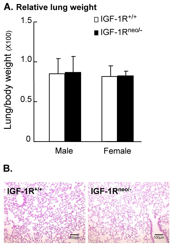
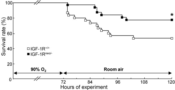
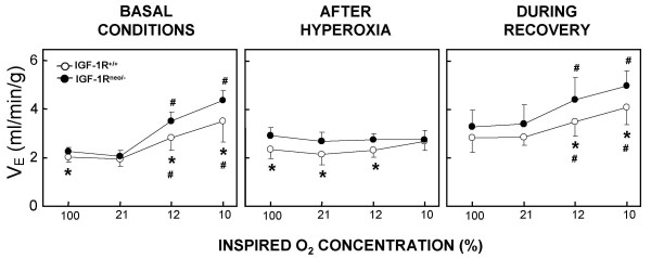
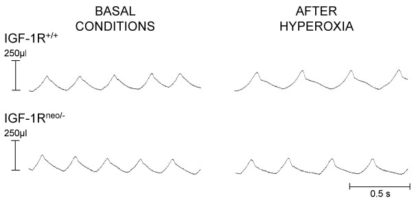
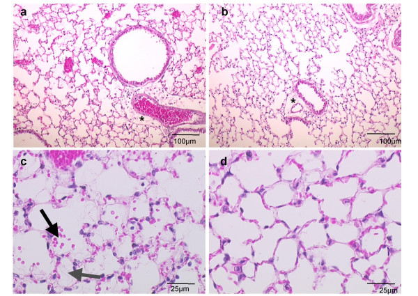
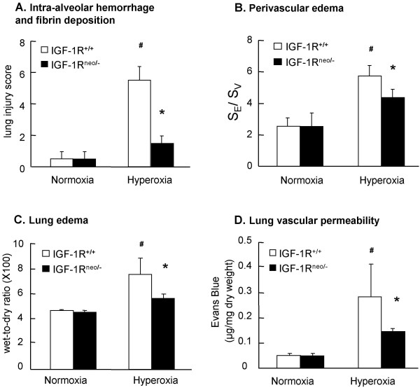
Results: Strikingly, after 72 h of exposure to 90% O2, IGF-1Rneo/- mice had a significantly better survival rate during recovery than IGF-1R+/+ mice (77% versus 53%, P < 0.05). The pulmonary injury was consistently, and significantly, milder in IGF-1Rneo/- mice which developed conspicuously less edema and vascular extravasation than controls. Also, hyperoxia-induced abnormal pattern of breathing which precipitated respiratory failure was elicited less frequently in the IGF-1Rneo/- mice.

Conclusion: Together, these data demonstrate that a decrease in IGF-1R signaling in mice protects against oxidant-induced lung injury.


Figures

Figure 1
Figure 1
Lung weight and histology under basal conditions. (A) Mean values (×100) of lung-to-body weight ratio (± SD) in IGF-1R+/+ mice (male: n = 11; female: n = 5) and IGF-1Rneo/- mice (male: n = 7; female: n = 4). (B) Representative histology of hematoxylin-eosin stained lung sections from IGF-1R+/+ and IGF-1Rneo/- mice. Magnification: 10× objective.
Figure 2
Figure 2
Survival rate following exposure to 90% oxygen. The survival times of IGF-1R+/+ mice (male: n = 15; female: n = 15) and IGF-1Rneo/- mice (male: n = 16; female: n = 15) were measured following exposure to 90% oxygen. Data are expressed as percentage of mice alive at each time point. The asterisk indicates significant differences between IGF-1R+/+ and IGF-1Rneo/- mice, as determined by Kaplan-Meier analysis; P < 0.05.
Figure 4
Figure 4
Ventilatory responses before and after hyperoxia, and during recovery. Minute ventilation (VE) in response to decreasing levels of inspired O2. In each one of the three conditions, mean values (± SD) of VE in IGF-1R+/+ and IGF-1Rneo/- mice were recorded under 90% O2, 21% O2 and at two levels of hypoxia (12% and 10% O2). Values with asterisks are significantly different from control values at 90% O2. Crosses indicate significant differences from normoxic control values, and asterisks indicate significant differences between IGF-1R+/+ and IGF-1Rneo/- mice; P < 0.05.
Figure 3
Figure 3
Respiratory pattern before and after hyperoxia. Typical respiratory pattern in a control IGF-1R+/+ mouse (upper traces) and in an IGF-1Rneo/- mouse (lower traces) under basal conditions and following hyperoxia. Expiratory pauses were observed in most control mice but not in IGF-1Rneo/- mice.
Figure 5
Figure 5
Histology following hyperoxic injury. Hematoxylin-eosin stained lung sections of (a) IGF-1R+/+, and (b) IGF-1Rneo/- mice following 72 h of hyperoxic exposure, illustrating perivascular and peribronchiolar edema as indicated by asterisks. Magnification: 10× objective. Representative histology of lung sections demonstrates (c) focal alveolar hemorrhages (black arrow) and hyaline membrane formation (gray arrow) in IGF-1R+/+ lungs, and (d) minimal lesions in IGF-1Rneo/- mice. Magnification: 40× objective.
Figure 6
Figure 6
Hyperoxia-induced lung injury, lung edema and vascular permeability. Parameters of lung damage were considered under basal conditions and following hyperoxia. (A) Lung injury scores from IGF-1R+/+ (n = 4) and IGF-1Rneo/- (n = 4) mice under basal conditions (normoxia) and under hyperoxia. A cumulative score was based on evaluation of hemorrhage and intraalveolar deposition (6 random sections per lung). (B) Quantitated perivascular edema from IGF-1R+/+ (n = 4) and IGF-1Rneo/- (n = 4) mice under basal conditions (normoxia) and under hyperoxia. Surface of perivascular edema was reported to the surface of the vessel, and noted SE/SV (5 selected sections per lung). (C) Lung edema was determined as mean values (± SD) of wet-to-dry lung weight ratio under basal conditions (normoxia) in IGF-1R+/+ (n = 4) and IGF-1Rneo/- (n = 4) mice, and under hyperoxia in IGF-1R+/+ (n = 7) and IGF-1Rneo/- (n = 8) mice. (D) Evans Blue dye extravasation was assessed as mean values (± SD) in IGF-1R+/+ (n = 13) and IGF-1Rneo/- (n = 11) mice. Crosses indicate significant differences between hyperoxic and normoxic values in IGF-1R+/+ mice, and asterisks indicate significant differences between IGF-1R+/+ and IGF-1Rneo/- mice under hyperoxic conditions; P < 0.05.

Similar articles

Cited by

References

    1. Lahiri S, Mokashi A, Shirahata M, Andronikou S. Chemical respiratory control in chronically hyperoxic cats. Respir Physiol. 1990;82:201–215. doi: 10.1016/0034-5687(90)90035-W. - DOI - PubMed
    1. Crapo JD, Barry BE, Foscue HA, Shelburne J. Structural and biochemical changes in rat lungs occurring during exposures to lethal and adaptive doses of oxygen. Am Rev Respir Dis. 1980;122:123–143. - PubMed
    1. Barazzone C, Horowitz S, Donati YR, Rodriguez I, Piguet PF. Oxygen toxicity in mouse lung: pathways to cell death. Am J Respir Cell Mol Biol. 1998;19:573–581. - PubMed
    1. Warner BB, Stuart LA, Papes RA, Wispe JR. Functional and pathological effects of prolonged hyperoxia in neonatal mice. Am J Physiol. 1998;275:L110–7. - PubMed
    1. Coalson JJ, Winter VT, Siler-Khodr T, Yoder BA. Neonatal chronic lung disease in extremely immature baboons. Am J Respir Crit Care Med. 1999;160:1333–1346. - PubMed

Publication types

LinkOut - more resources