Comparative Study
. 2005 Apr 8:6:24.
doi: 10.1186/1471-2202-6-24.

Hsp27 and axonal growth in adult sensory neurons in vitro

Affiliations
Comparative Study

Hsp27 and axonal growth in adult sensory neurons in vitro

Kristy L Williams et al. BMC Neurosci. .

Abstract

Background: Neurite growth can be elicited by growth factors and interactions with extracellular matrix molecules like laminin. Among the targets of the signalling pathways activated by these stimuli are cytoskeletal elements, such as actin, tubulin and neurofilaments. The cytoskeleton can also be modulated by other proteins, such as the small heat shock protein Hsp27. Hsp27 interacts with actin and tubulin in non-neuronal cells and while it has been suggested to play a role in the response of some neurons to injury, there have been no direct studies of its contribution to axonal regeneration.

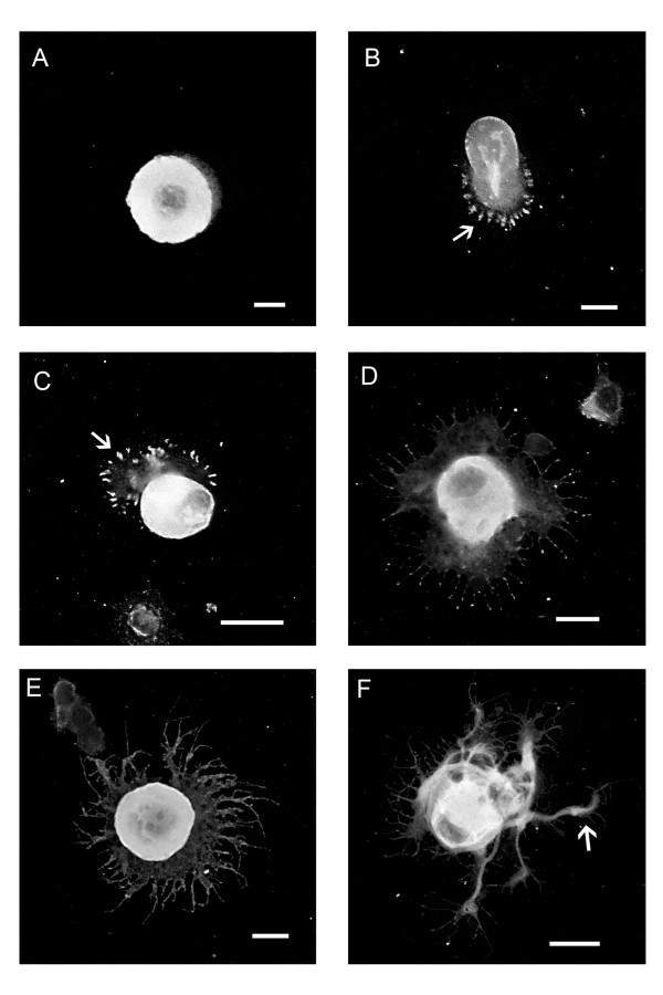
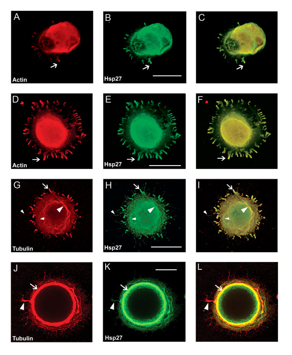
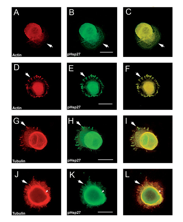
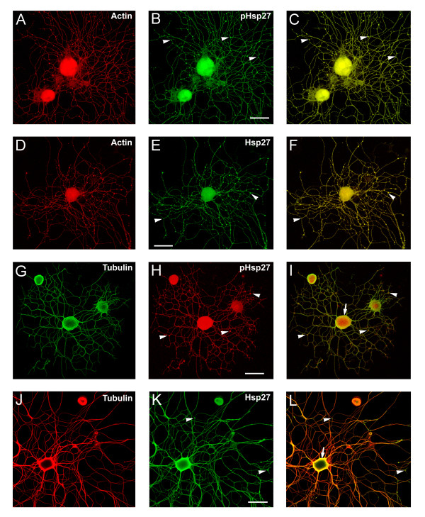
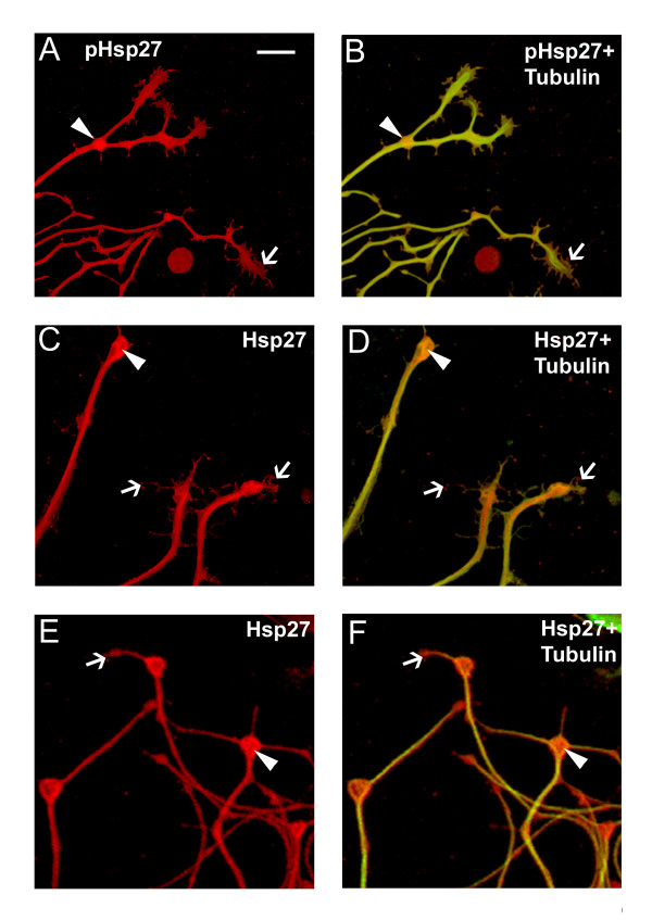
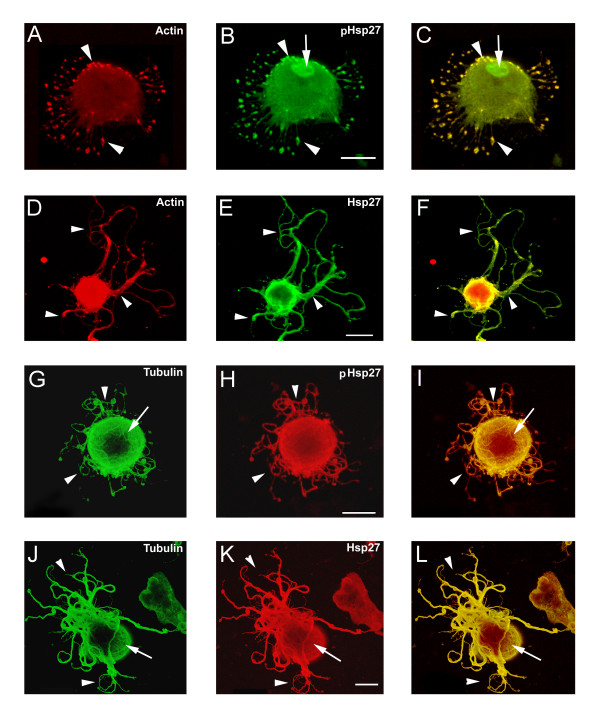
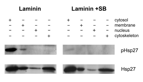
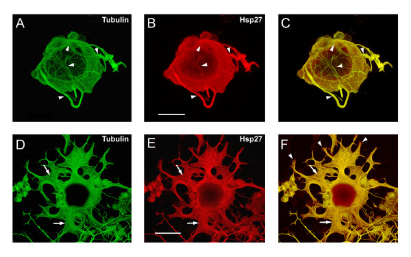
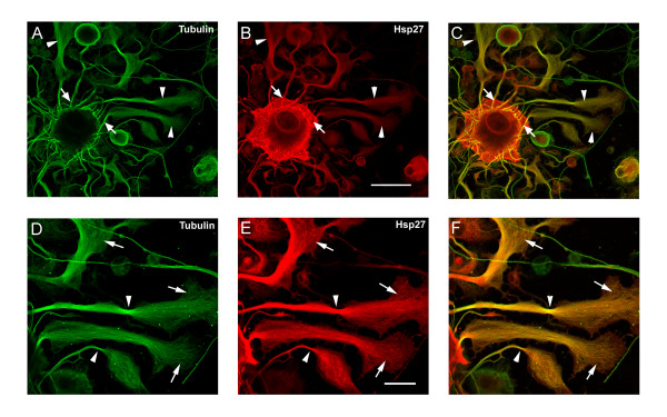
Results: We have investigated neurite initiation and process extension using cultures of adult dorsal root ganglion (DRG) sensory neurons and a laminin stimulation paradigm. Employing confocal microscopy and biochemical analyses we have examined localization of Hsp27 at early and later stages of neurite growth. Our results show that Hsp27 is colocalized with actin and tubulin in lamellopodia, filopodia, focal contacts and mature neurites and growth cones. Disruption of the actin cytoskeleton with cytochalasin D results in aberrant neurite initiation and extension, effects which may be attributable to alterations in actin polymerization states. Inhibition of Hsp27 phosphorylation in our cultures results in an atypical growth pattern that may be attributable to an effect of pHsp27 on the stability of the actin cytoskeleton.

Conclusion: We observed colocalization of the phosphorylated and non-phosphorylated forms of Hsp27 with actin and tubulin in both very early and later stages of neurite growth from cultured adult DRG neurons. The colocalization of Hsp27 and pHsp27 with actin in lamellopodia and focal contacts at early stages of neurite growth, and in processes, branch points and growth cones at later stages, suggests that Hsp27 may play a role in neuritogenesis and subsequent neurite extension, and potentially in the patterning of this growth. Hsp27 has been reported to play a key role in modulating actin cytoskeletal dynamics as an actin-capping protein in non-neuronal cells. Our results suggest that this may also be the case in neurons and support a role for Hsp27 in neurite outgrowth via its phosphorylation state-dependent interactions with actin.


Figures

Figure 1
Figure 1
Laminin stimulation elicits lamellopodia and process formation in adult sensory neurons. DRG neurons plated on polylysine were stimulated with laminin in solution for 5 min, 15 min, 30 min, 1 hr, 6 hrs, 24 hrs. After fixation, neurons were stained with rhodamine-phalloidin to detect actin and images obtained using confocal microscopy. Panels A-F provide representative examples of the various stages of lamellopodia formation and eventual process protrusion, and show various distinctive stages in neuronal membrane expansion and neurite growth. At the earliest stages, lamellopodia are formed (A- 5 min, B- 15 min, C- 30 min) with evidence of focal contacts (arrows) at the leading edge of the lamellopodia (B, C). In D (1 hr) and E (6 hrs) filopodia begin to protrude from the lamellopodium around the circumference of the neuron. Eventually, these processes appear to coalesce into one or more neurites that continue to extend (F- 24 hrs, arrow). Scale bar – 20 μm.
Figure 2
Figure 2
Hsp27 co-localizes with actin and tubulin at early stages of neurite growth. Neurons were plated on polylysine and stimulated with laminin for 1–6 hrs. Following fixation, neurons were labelled with rhodamine-phalloidin (red-A, D) or immunostained with antibodies directed against total tubulin (red – G, J) or Hsp27 (green -B, E, H, K). Images were obtained with confocal microscopy and panels C, F, I, L represent the merged images of the single channel images. Note colocalization of Hsp27 and actin in the lamellopodium (A-C, arrow) and in focal contacts observed in D-F (arrow). In panels G-I, there is some colocalization of the staining for tubulin and Hsp27 in the cortical region (large arrowhead) and in small processes emerging from the soma (arrow). In panels J-L, there is a more distinct colocalization of tubulin and Hsp27 in the cortical area (arrow, J-L) as well as in an obvious process that seems to be wrapping around the cell and finally extending (arrowhead, J-L). Scale bar – 20 μm
Figure 3
Figure 3
pHsp27 also co-localizes with actin and tubulin at early stages of neurite growth. Neurons plated on polylysine and stimulated with laminin for 1–6 hrs were also immunostained with antibodies directed against phosphorylated Hsp27 (pHsp27Ser15) to examine colocalization with actin or tubulin. A, D – rhodamine-phalloidin (red); B, E, H, K – pHsp27 (green); G, J – tubulin (red). The respective merged images are presented in panels C, F, I, L. Actin and pHsp27 appear to be colocalized in the body of the lamellopodium in A-C, but actin seems to be excluded from the leading edge (arrows). There is also localization of pHsp27 and actin in focal contacts (D-F, arrows). pHsp27 also colocalizes with tubulin in focal contacts (G-I, arrow), in a cortical ring (arrowhead) and in processes emerging from the cell body (J-L, arrow). Scale bar – 20 μm.
Figure 4
Figure 4
Hsp27 continues to be expressed and localized with cytoskeletal elements in neurons and neuritic networks. In these experiments, neurons were plated on laminin (no added neurotrophins) and fixed 24 hrs after plating. As shown in the images, many neurons exhibit extensive neuritic growth under these conditions. A-F: Neurons were labelled with rhodamine-phalloidin (A, D, red), and immunostained for pHsp27 (B, green) and Hsp27 (E, green); C, F – merged images. G-L: Neurons were immunostained for tubulin (G, green; J, red), pHsp27 (H, red), Hsp27 (K, green); I, L – merged images. Hsp27 and pHsp27 are expressed throughout the neuritic network, and there is colocalization of these with actin (C, F) and less so with tubulin (I, L). Note the accumulation of pHsp27 and Hsp27 at point of branching of neurites (arrowheads- B, C, E, F, H, I, K, L). The cortical colocalization of tubulin with pHsp27 and Hsp27 is still evident at this stage of neurite growth (arrows – I, L). Scale bar – 50 μm.
Figure 5
Figure 5
Co-localization of Hsp27 and actin in growth cones of growing neurites. Growth cones from neurons plated on laminin as outlined for Figure 4 were observed to express both pHsp27 and Hsp27. pHsp27 (A) and Hsp27 (C, E, red), shown together with tubulin (green-yellow) in the merged images (B, D, F), are present in growth cones and filopodia extending from the growth cones (arrows). There is also an accumulation in the core of growth cones and at points of neurite branching (arrowheads- A-F). Note that the tubulin staining does not completely overlap with pHsp27 or Hsp27, particularly in some of the extending filopodia (B, arrow) and the core of the growth cones in D, F (arrowheads). Scale bar – 10 mm.
Figure 6
Figure 6
Disruption of the actin cytoskeleton results in aberrant neurite growth. Neurons plated on LN were treated with cytochalasin D (2 mM, added 3 hrs after plating), fixed 24 hrs later and stained for pHsp27 (B, H), Hsp27 (E, K), actin (A, D) or tubulin (G, J). The respective merged images are presented in panels C, F, I and L. The cytochalasin D treatment resulted in various atypical patterns of growth. One phenotype was the elaboration of numerous processes or microspikes as seen in panels A-C, with obvious accumulation of actin and pHsp27 especially at the tips of the microspikes (arrowheads); pHsp27, but not actin, accumulates in the nucleus (B, C, arrow). Abnormal process extension was also observed. In the neuron shown in D-F, some extension was observed although there was now less colocalization of actin with the Hsp27 (arrowheads). Panels G-L show tubulin staining along with either pHsp27 or Hsp27; the nature of the cytoskeletal network is clearer in these examples (arrows). Arrowheads point to atypical neurite growth, eg., lacking the usual radial branching pattern as seen in Fig 4. Scale bar – 20 mm.
Figure 7
Figure 7
p38 MAPK inhibition blocks phosphorylation of Hsp27. Neurons plated on laminin were exposed to p38 MAPK inhibitors, SB203580 and SB202190 (10 mM each). Cells were sampled at 24 hrs post SB addition, using cellular subfractionation (as described in the Methods). The resulting protein from cytosol, membrane, nucleus and cytoskeleton fractions was electrophoresed and the blot subsequently probed for pHsp27 and Hsp27. Inhibition of p38 MAPK activity (laminin+SB) results in attenuation of the Hsp27 phosphorylation.
Figure 8
Figure 8
Aberrant neurite growth following inhibition of Hsp27 phosphorylation. Neurons plated on laminin were treated with p38 MAPK inhibitors (SB203580 and SB202190, 10 μM each, added 3 hrs after plating) and fixed 24 hrs later. Representative results are presented. Some neurons showed abortive extension, with neurites wrapping around the cell body, such as the example in panels A-C (arrowheads, A, tubulin, B, Hsp27, C, merged image). In another example, numerous processes were observed, but these terminated in large, flattened and splayed growth cones, as shown in panel D-F (D, tubulin, E, Hsp27, F, merged image). The fibrillar nature of the Hsp27 (E, arrows) and tubulin (D, arrows) is evident and the sites of colocalization with tubulin are also apparent (F, arrows). Also note that there is not a complete overlap of Hsp27 and tubulin at the tips of the growth cones (F, arrowheads). Scale bar – 20 μm.
Figure 9
Figure 9
Flattened growth cones and processes show co-localization of tubulin and Hsp27. This figure shows another example of a neuron treated with the p38 MAPK inhibitors as outlined in Figure 8. Colocalization of Hsp27 (B, red) with tubulin (A, green) is apparent in the emerging processes (C, arrows), and in the flattened and splayed growth cones (A-C, arrowheads); scale bar – 50 μm. At a higher magnification (D-F), loss of microtubule bundling is observed (arrowheads) along with the fibrillar nature of Hsp27 and colocalization with tubulin (arrows); scale bar – 20 μm.

Similar articles

Cited by

References

    1. Tucker BA, Rahimtula M, Mearow KM. Integrin activation and neurotrophin signaling cooperate to enhance neurite outgrowth in sensory neurons. J Comp Neurol. 2005;486 - PubMed
    1. da Silva JS, Dotti CG. Breaking the neuronal sphere: regulation of the actin cytoskeleton in neuritogenesis. Nat Rev Neurosci. 2002;3:694–704. doi: 10.1038/nrn918. - DOI - PubMed
    1. Dehmelt L, Smart FM, Ozer RS, Halpain S. The role of microtubule-associated protein 2c in the reorganization of microtubules and lamellipodia during neurite initiation. J Neurosci. 2003;23:9479–9490. - PMC - PubMed
    1. Dehmelt L, Halpain S. Actin and microtubules in neurite initiation: are MAPs the missing link? J Neurobiol. 2004;58:18–33. doi: 10.1002/neu.10284. - DOI - PubMed
    1. Charette SJ, Landry J. The interaction of HSP27 with Daxx identifies a potential regulatory role of HSP27 in Fas-induced apoptosis. Ann N Y Acad Sci. 2000;926:126–131. - PubMed

Publication types

MeSH terms