. 2005 May;23(2-3):341-53.
doi: 10.1016/j.cogbrainres.2004.11.014.

Event-related potentials reveal dissociable mechanisms for orienting and focusing visuospatial attention

Affiliations

Event-related potentials reveal dissociable mechanisms for orienting and focusing visuospatial attention

Shimin Fu et al. Brain Res Cogn Brain Res. 2005 May.

Abstract

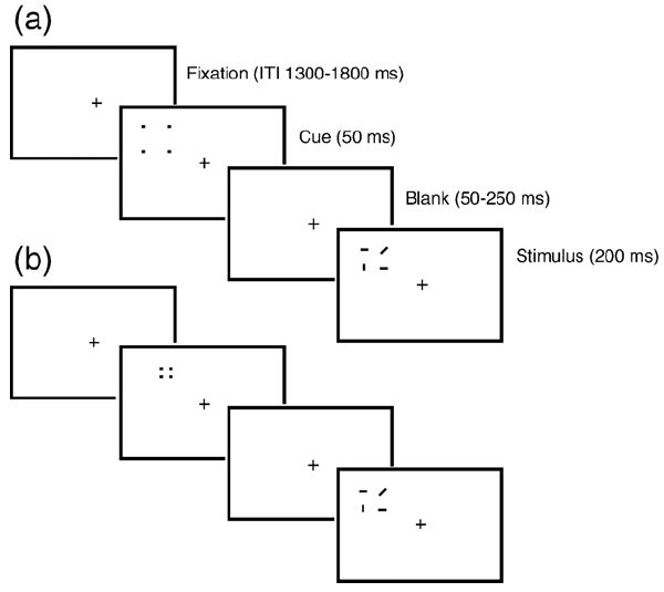
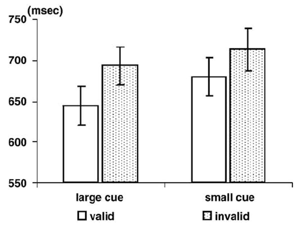
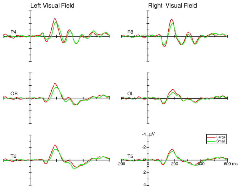
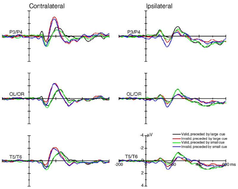
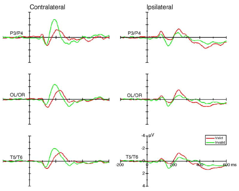
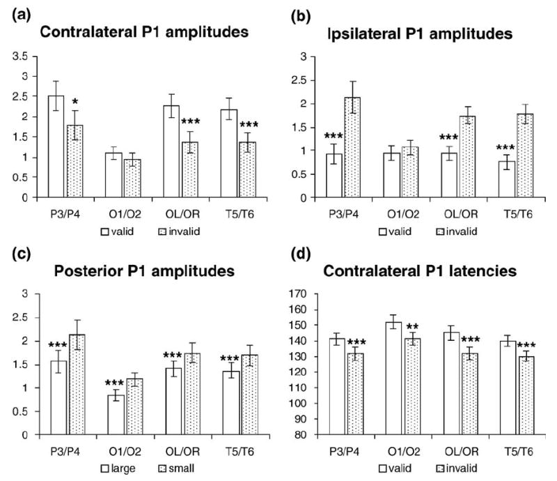
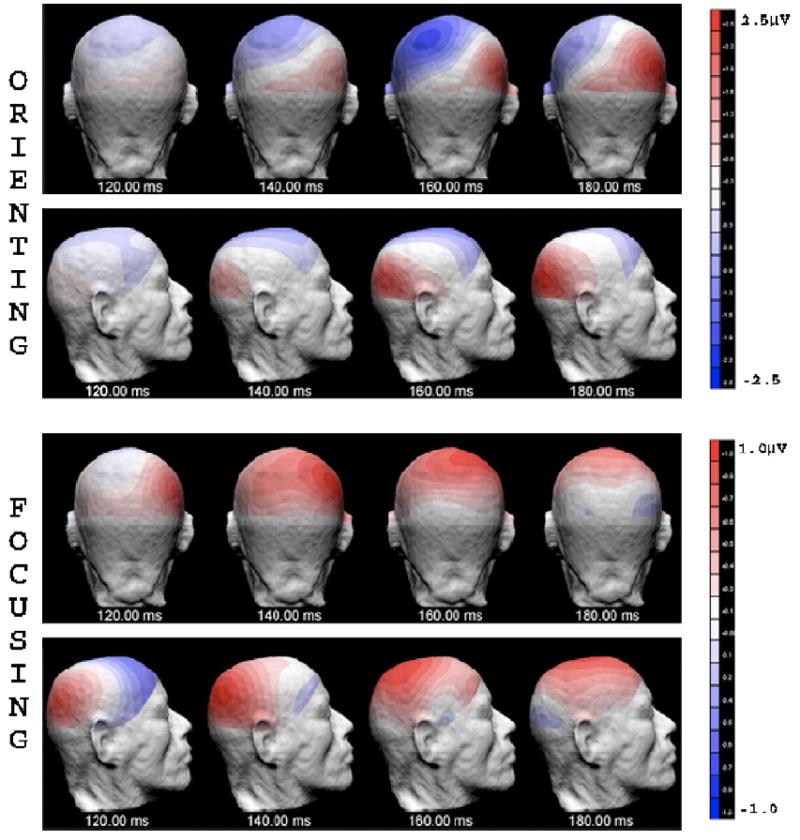
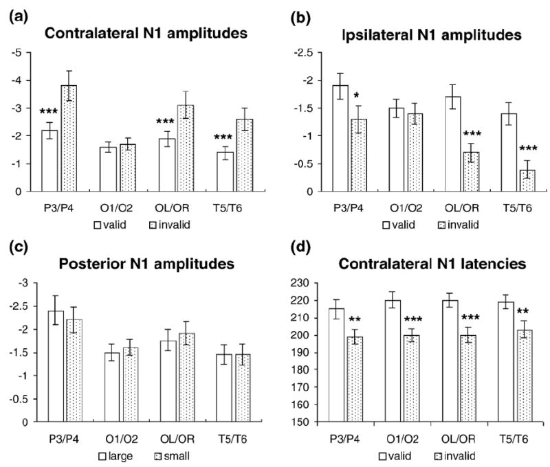
The neural mechanisms supporting visuospatial orienting and focusing were investigated by recording event-related potentials (ERPs) in a cued, line-orientation discrimination task. Search arrays flashed randomly in the left or right visual field and were preceded by peripheral cues that varied in validity (valid or invalid, with 50% each) and size (large or small, with 50% each). Facilitation of response time was observed for valid trials, regardless of cue size. In contrast to previous cued search studies, however, small (i.e., more precise) cues were associated with delayed responses. Both the timing and the amplitudes of the early ERP components, P1 and N1, were modulated by attentional orienting, with valid trials eliciting a larger and later contralateral vP1 (ventral P1) and a smaller and later contralateral N1 compared to invalid trials. Attentional focusing modulated only the amplitudes of the P1 component, with precisely cued trials eliciting a larger dP1 (dorsal P1) than less precisely cued trials at both contralateral and ipsilateral sites. Thus, both attentional orienting and focusing modulate early stimulus processing stages that overlap in time, but with dissociable effects on the scalp distribution of these components, indicating possibly different underlying mechanisms. In addition, the results support the notion that voluntary and involuntary allocations of visuospatial attention are mediated by different underlying neural processes.


Figures

Fig. 1
Fig. 1
Schematic illustration of the procedure for the present study. The cues consisted of four small dots that formed an imaginary rectangle. The size of the cues could be large (a) or small (b). The search array consisted of two horizontal lines, one vertical line and one diagonal line. The large valid cues covered the whole search array (a), whereas the small valid cues covered only the quadrant containing the diagonal line (b). On 50% of trials, the search array appeared at the same side as the cue. Cues and search arrays could appear either to the left or to the right of fixation with equal probability. Invalid trials are not shown here; however, they were identical to valid trials except that the cue and the search array appeared on opposite sides of the screen from one another.
Fig. 2
Fig. 2
The mean reaction times (RTs) and standard errors for the targets as a function of cue validity and cue size.
Fig. 3
Fig. 3
The grand average of ERPs (across 16 subjects) elicited by the large (red lines) and small cues (green lines) at the posterior contralateral sites when the cues appeared in the left (left column) and right (right column) visual field. Data were averaged across valid and invalid cues.
Fig. 4
Fig. 4
The grand average of ERPs (across 16 subjects) elicited by standard stimuli at the posterior contralateral (left column) and ipsilateral (right column) sites. Data were averaged across visual field and hemisphere.
Fig. 5
Fig. 5
The grand average of ERPs (across 16 subjects) elicited by the valid (red lines) and invalid (green lines) stimuli at the contralateral (left column) and ipsilateral (right column) recording sites. Data were averaged across visual field, hemisphere, and cue size.
Fig. 6
Fig. 6
The mean voltages and peak latencies of the cue validity effects and cue size effects on the P1 components, along with the statistical results (*P < 0.05; **P < 0.005; ***P < 0.001). (a) The cue validity effects on the amplitude of contralateral P1; (b) the cue validity effects on the amplitude of ipsilateral P1; (c) the cue size effects on the amplitude of posterior P1; (d) the cue validity effects on the latency of contralateral P1.
Fig. 7
Fig. 7
The 3D scalp voltage distribution of the orienting-related ERP (cue validity effect, obtained by subtracting ERPs of the invalid condition from ERPs of the valid condition), and the focusing-related ERP components (cue size effect, obtained by subtracting ERPs of the large valid cue condition from ERPs of the small valid cue condition), when the target array appeared in the left visual field. Data for orienting were averaged across small cue and large cue conditions. Only the ERPs of valid trials for the small and large cue conditions were used regarding the subtraction for focusing-related component. Data are shown in back view and right view of the head, every 20 ms from 120 to 180 ms after the onset of search array. Note that the focusing-related ERP had a more dorsal distribution as compared with a more ventral distribution of the orienting-related ERP at 140 ms (the P1 time range).
Fig. 8
Fig. 8
The grand average of ERPs (across 16 subjects) elicited by stimuli that were preceded by large cues (red lines) and by small cues (green lines) at the posterior contralateral (left column) and ipsilateral recording sites (right column). Data were averaged across visual field, hemisphere, and cue validity.
Fig. 9
Fig. 9
The mean voltages and peak latencies of the cue validity effects and cue size effects on the N1 component, along with the statistical results (*P < 0.05; **P < 0.005; ***P < 0.001). (a) The cue validity effects on the amplitude of contralateral N1; (b) the cue validity effects on the amplitude of ipsilateral N1; (c) the cue size effects on the amplitude of posterior N1; (d) the cue validity effects on the latency of contralateral N1.

References

    1. Anllo-Vento L, Hillyard SA. Selective attention to the color and direction of moving stimuli: electrophysiological correlates of hierarchical feature selection. Percept Psychophys. 1996;58:191–206. - PubMed
    1. Carrasco M, Yeshurun Y. The contribution of covert attention to the set-size and eccentricity effects in visual search. J Exp Psychol Hum Percept Perform. 1998;24:673–692. - PubMed
    1. Castiello U, Umilta C. Size of the attentional focus and efficiency of processing. Acta Psychol (Amsterdam) 1990;73:195–205. - PubMed
    1. Castiello U, Umilta C. Splitting focal attention. J Exp Psychol Hum Percept Perform. 1992;18:837–848. - PubMed
    1. Cheal M, Lyon DR. Central and peripheral precuing of forced-choiced discrimination. Q J Exp Psychol, A. 1991;43:859–880. - PubMed

Publication types

LinkOut - more resources