. 2005 Apr 8;121(1):101-13.
doi: 10.1016/j.cell.2005.01.035.

Ubiquitin ligase Smurf1 controls osteoblast activity and bone homeostasis by targeting MEKK2 for degradation

Affiliations

Ubiquitin ligase Smurf1 controls osteoblast activity and bone homeostasis by targeting MEKK2 for degradation

Motozo Yamashita et al. Cell. .

Abstract

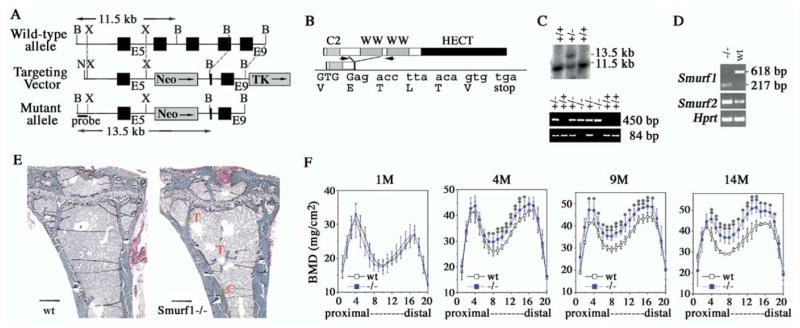
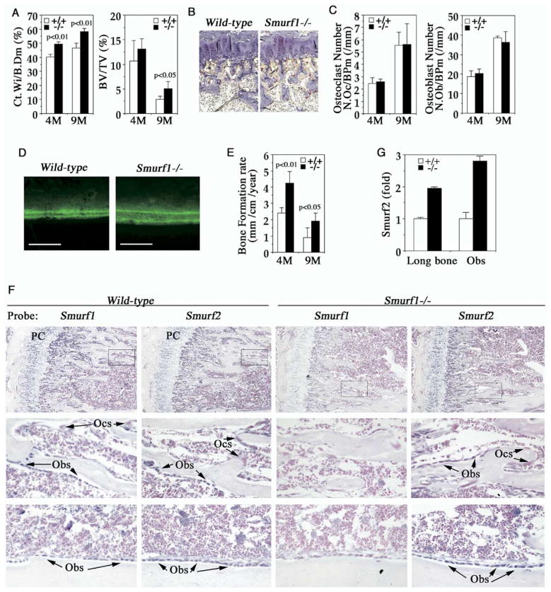
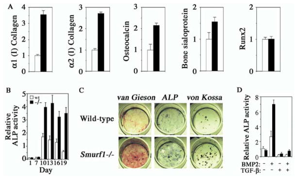
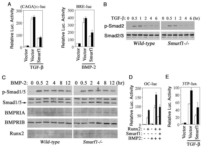
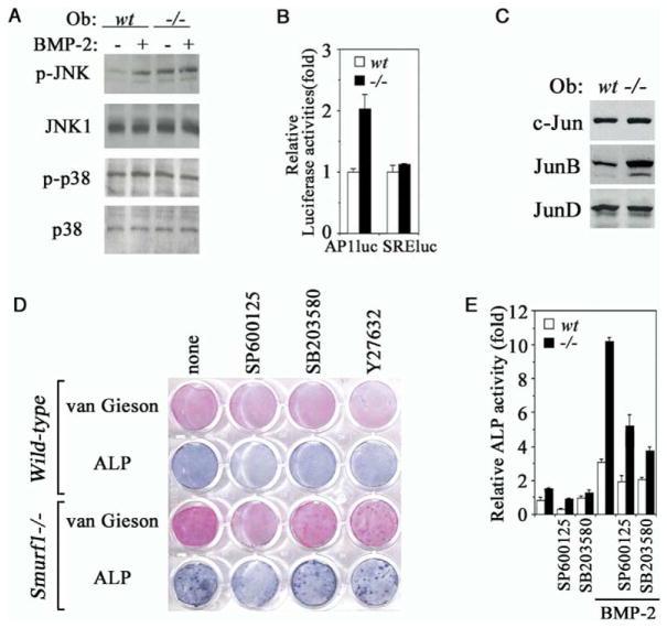
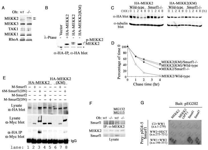
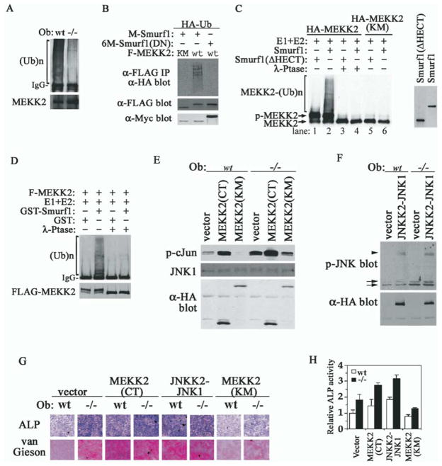
Bone is constantly resorbed and formed throughout life by coordinated actions of osteoclasts and osteoblasts. Here we show that Smurf1, a HECT domain ubiquitin ligase, has a specific physiological role in suppressing the osteogenic activity of osteoblasts. Smurf1-deficient mice are born normal but exhibit an age-dependent increase of bone mass. The cause of this increase can be traced to enhanced activities of osteoblasts, which become sensitized to bone morphogenesis protein (BMP) in the absence of Smurf1. However, loss of Smurf1 does not affect the canonical Smad-mediated intracellular TGFbeta or BMP signaling; instead, it leads to accumulation of phosphorylated MEKK2 and activation of the downstream JNK signaling cascade. We demonstrate that Smurf1 physically interacts with MEKK2 and promotes the ubiquitination and turnover of MEKK2. These results indicate that Smurf1 negatively regulates osteoblast activity and response to BMP through controlling MEKK2 degradation.


Figures

Figure 1
Figure 1. Increased Bone Mass in Smurf1−/− Mice
(A) Generation of Smurf1-deficient mice. Closed box, exon; shaded box, selective marker, pGKneo or pGKtk; N, NotI; X, XhoI; B, BamHI. (B) Structural organization of Smurf1 protein. Sequence surrounding the acquired splicing junction between Exons 5 (upper case) and Exon 9 (lower case) was confirmed by sequencing the RT-PCR products in (D). (C) (Upper panel) Southern blot analysis of the homologous recombination using the 5′ external probe (shown in [A]). Wt, 11.5 kb; mutant, 13.5 kb. (Lower panel) PCR genotyping. Wt and mutated alleles yield 84 bp and 450 bp products, respectively. (D) RT-PCR analyses of Smurf1 and Smurf2 expression using total RNA isolated from newborn wild-type and Smurf1−/− mice. For Smurf1 expression, a primer pair from E5 and E9 (arrows in [B]) was used. Amplification of Hprt was used as an internal control. (E) Villanueva Goldner staining of plastic sections of tibiae from mice at age of 9 months. Scale bar, 0.4 mm. C, cortical bone; T, trabecular bone. (F) BMD measurement in 20 divisions from the proximal to distal femora of male mice at ages of 1, 4, 9, and 14 months. Wt: n = 4; Smurf1−/−: n = 8 (1 month old), or n = 6 (4, 9, 14 month old). Statistical difference of Smurf1−/− mice from Wt mice was assessed by Student’s t test. *p < 0.05, **p < 0.01.
Figure 2
Figure 2. Enhanced Osteoblast Activity in Smurf1−/− Mice
(A) Bone morphometric analyses of 4- and 9-month-old male mice (n = 4). Ct.Wi/B. Dm, cortex bone width per bone diameter; BV/TV, trabecular bone volume per tissue volume. Statistical difference between two mice groups was assessed by Student’s t test. (B) TRAP staining of osteoclasts in proximal epiphysis of tibia from 4-month-old mice. (C) Quantification of osteoclast (left) and osteoblast (right) cell numbers. (D) Calcein-labeled mineralization fronts in tibiae cortex bone from 4-month-old mice by fluorescent micrography. Scale bar = 0.2 mm. (E) Quantification of bone formation rate measured with calcein double-labeling. (F) In situ hybridization analyses of Smurf1 and Smurf2 expression in tibia at the metaphysis (top panels) and near cortical bone (bottom panels). (Middle panels) Insets of top panels at higher magnification. Sections were counterstained with nuclear fast red. PC, proliferative chondrocytes; Obs, osteoblasts; Ocs, osteoclasts. (G) Real-time RT-PCR measurement of Smurf2 transcript from lone bones or osteoblasts.
Figure 3
Figure 3. Enhanced ECM Production in Smurf1−/− Mice and BMP Sensitivity of Smurf1−/− Osteoblasts
(A) Real-time PCR of osteoblast marker genes from long bone mRNA of 4-month-old wild-type (open column) or Smurf1−/− mice (closed column). Values are presented as relative expression. (B) Time course of ALP activity in differentiating calvaria cell culture. (C) Calvaria-derived osteoblasts were differentiated for 12 days in vitro and stained for collagen matrix formation (van Gieson) and ALP activity. Von Kossa staining for mineralized nodules was done after culturing for 21 days. (D) ALP activity of calvaria-derived cells after 7 days of differentiation in the presence of exogenous BMP-2 and/or TGFβ.
Figure 4
Figure 4. Normal Smad-Dependent TGFβ and BMP Response in Smurf1−/− Osteoblasts
(A) Smad-dependent TGFβ or BMP signaling in osteoblasts measured by (CAGA)12-Luc or BRE-luc transcription reporter assay. (B) Western analyses of TGFβ-induced Smad2 phosphorylation and the steady-state levels of the endogenous Smad2 and Smad3. (C) Western analyses of BMP-2-induced Smad1/5 phosphorylation and the steady-state levels of the endogenous total Smad1/5, BMP receptors (BMPRIA and BMPRIB), and Runx2. (D) Effect of loss of Smurf1 on transcription from the Runx2-dependent osteocalcin promoter (OC-Luc). (E) Effect of loss of Smurf1 on transcription from 3TP-Lux. Open bar, wild-type; closed bar, Smurf1−/−.
Figure 5
Figure 5. Elevated JNK Activity in Smurf1−/−Osteoblasts
(A) Western analyses of JNK and p38 MAPK phosphorylation in isolated osteoblasts. Where indicated, BMP-2 was added at 100 ng/ml for 30 min prior to sample preparation. (B) JNK-dependent AP-1 transcription as measured by AP-1-luc reporter. SRE-luc, a control reporter containing the serum response element. (C) Western analyses of endogenous c-Jun, JunB, and JunD expression. (D) Blocking the augmented collagen matrix production and ALP activity by JNK inhibitor in Smurf1−/− osteoblasts. Experiments were carried out as in Figure 3C except for the addition of SP600125, SB203580, or Y27632. (E) ALP quantification in osteoblasts cultured for 12 days. The relative activity was expressed as fold to the ALP activity of wild-type osteoblasts in the absence of inhibitors and BMP-2.
Figure 6
Figure 6. Accumulation of Phosphorylated MEKK2 in Smurf1−/− Osteoblasts and Physical Interaction between Smurf1 and MEKK2
(A) Western analyses of MEKK2, MEKK3, MEKK1, TAK1, and RhoA in osteoblasts. (B) Western analyses of HA-tagged MEKK2 or kinase-deficient MEKK2 (KM) after immunoprecipitation from transfected Smurf1−/− MEFs. (C) Western analyses of HA-MEKK2 and HA-MEKK2(KM) in transfected MEFs at the indicated intervals following cycloheximide treatment. (D) [35S]-methionine labeling and chase studies of MEKK2 and MEKK2 (KM) in wild-type Smurf1−/− MEFs. The amount of each labeled protein was expressed as the percentage of that at the beginning of chase (time 0). (E) Interaction between exogenous Smurfs and MEKK2 in Smurf1−/− MEFs. Expression of the HA-tagged MEKK2s and the Myc-tagged Smurfs was shown in top and middle panel, respectively. Only Smurf1(DN) coimmunoprecipitated with the wild-type MEKK2 and weakly with MEKK2(KM) (bottom panel, lanes 3 and 8). (F) Interaction between endogenous Smurf1 and MEKK2. MEKK2 was precipitated from wild-type and Smurf1−/− osteoblasts that were pre-treated with proteasome inhibitors and subjected to Western analysis with anti-Smurf1 antibody (top panel). The amount of total MEKK2 and Smurf1 present in cell lysates was shown at two bottom panels. (G) Requirements for direct interaction between Smurf1 and MEKK2 as revealed by yeast two-hybrid assays. Binding of the WW domains of Smurf1 to Smad1 or Smad7 was included as positive controls.
Figure 7
Figure 7. MEKK2 Is a Substrate of Smurf1-Mediated Ubiquitination and Activation of MEKK2-JNK Pathway Is Sufficient to Enhance Osteoblast Activity
(A) Ubiquitination of endogenous MEKK2 in osteoblasts. Polyubiquitinated MEKK2 was detected by anti-ubiquitin Western analysis after precipitation of MEKK2 from cells that were pretreated with proteasome inhibitors. (B) Smurf1 promotes MEKK2 ubiquitination. Ubiquitinated F-MEKK2 in Smurf1−/− MEFs was visualized by anti-HA-ubiquitin Western analysis after anti-FLAG immunoprecipitation. Expression of Myc-tagged Smurfs is shown in bottom panel. (C) In vitro polyubiquitination of MEKK2 by Smurf1 detected by anti-HA blot. HA-MEKK2 and HA-MEKK2(KM) were isolated by anti-HA antibody from transfected Hep3B cells. Myc-tagged Smurf1 and Smurf1(ΔHECT) were in vitro translated and are shown in the right panel. (D) In vitro ubiquitination of MEKK2 by purified GST-Smurf1 detected by anti-ubiquitin blot. FLAG-MEKK2 purified from transfected Drosophila S2 cells is shown at bottom panel. (E) In vitro kinase assay of JNK1 after immunoprecipitation from osteoblasts expressing MEKK2 mutants. GST-c-Jun was used as the kinase substrate and was analyzed by anti-p-c-Jun blot. Total JNK1, HA-MEKK2(CT), and MEKK2(KM) are shown below. (F) Western analyses of phosphorylated JNK in osteoblasts expressing HA-JNKK2-JNK1, which does not affect endogenous JNK activity. Arrows, endogenous p-JNK1/2; arrowhead, p-JNKK2-JNK1 fusion. (G) Ectopic expression of activated MEKK2 and JNK1 is sufficient to enhance osteoblast activity. Staining for ALP activity and collagen matrix was carried out after incubating in differentiation medium for 16 days. (H) ALP quantification of cells in (G) after differentiation for 12 days.

Comment in

References

    1. Alliston T, Choy L, Ducy P, Karsenty G, Derynck R. TGF-β-induced repression of CBFA1 by Smad3 decreases cbfa1 and osteocalcin expression and inhibits osteoblast differentiation. EMBO J. 2001;20:2254–2272. - PMC - PubMed
    1. Bakin AV, Rinehart C, Tomlinson AK, Arteaga CL. p38 mitogen-activated protein kinase is required for TGFβ-mediated fibroblastic transdifferentiation and cell migration. J Cell Sci. 2002;115:3193–3206. - PubMed
    1. Balemans W, Van Hul W. Extracellular regulation of BMP signaling in vertebrates: a cocktail of modulators. Dev Biol. 2002;250:231–250. - PubMed
    1. Bhargava U, Bar-Lev M, Bellows CG, Aubin JE. Ultrastructural analysis of bone nodules formed in vitro by isolated fetal rat calvaria cells. Bone. 1988;9:155–163. - PubMed
    1. Brown JD, DiChiara MR, Anderson KR, Gimbrone MA, Jr, Topper JN. MEKK-1, a component of the stress (stress-activated protein kinase/c-Jun N-terminal kinase) pathway, can selectively activate Smad2-mediated transcriptional activation in endothelial cells. J Biol Chem. 1999;274:8797–8805. - PubMed

MeSH terms

Substances