Comparative Study
. 2005 Apr 12;15(7):603-15.
doi: 10.1016/j.cub.2005.02.059.

NMDA receptors mediate olfactory learning and memory in Drosophila

Affiliations
Comparative Study

NMDA receptors mediate olfactory learning and memory in Drosophila

Shouzhen Xia et al. Curr Biol. .

Abstract

Background: Molecular and electrophysiological properties of NMDARs suggest that they may be the Hebbian "coincidence detectors" hypothesized to underlie associative learning. Because of the nonspecificity of drugs that modulate NMDAR function or the relatively chronic genetic manipulations of various NMDAR subunits from mammalian studies, conclusive evidence for such an acute role for NMDARs in adult behavioral plasticity, however, is lacking. Moreover, a role for NMDARs in memory consolidation remains controversial.

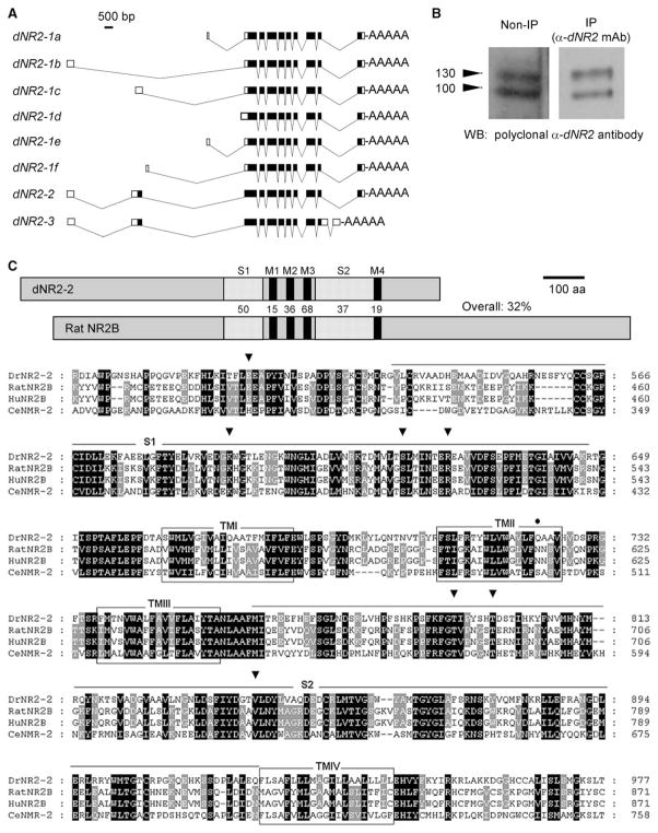
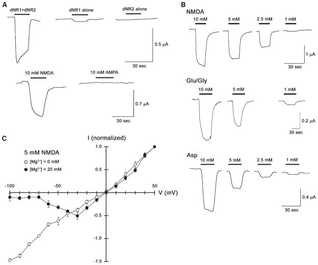
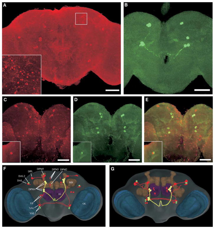
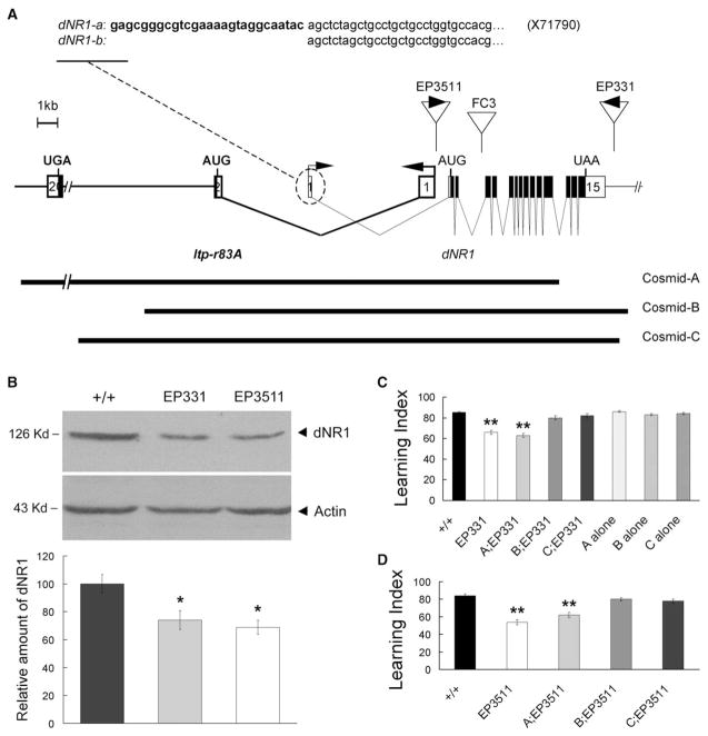
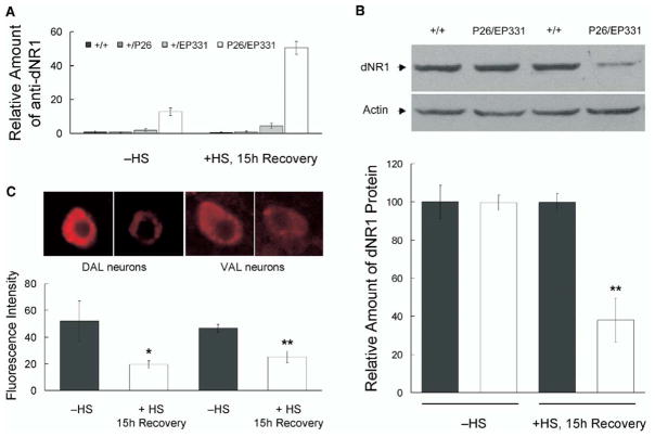
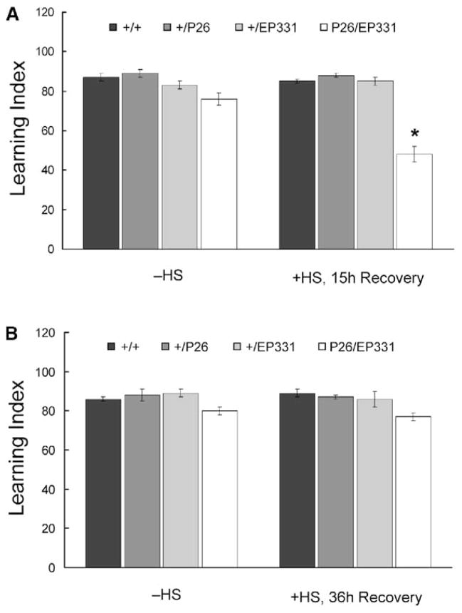
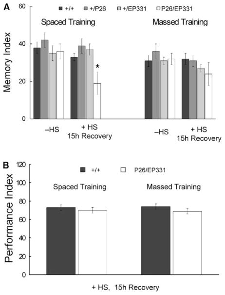
Results: The Drosophila genome encodes two NMDAR homologs, dNR1 and dNR2. When coexpressed in Xenopus oocytes or Drosophila S2 cells, dNR1 and dNR2 form functional NMDARs with several of the distinguishing molecular properties observed for vertebrate NMDARs, including voltage/Mg(2+)-dependent activation by glutamate. Both proteins are weakly expressed throughout the entire brain but show preferential expression in several neurons surrounding the dendritic region of the mushroom bodies. Hypomorphic mutations of the essential dNR1 gene disrupt olfactory learning, and this learning defect is rescued with wild-type transgenes. Importantly, we show that Pavlovian learning is disrupted in adults within 15 hr after transient induction of a dNR1 antisense RNA transgene. Extended training is sufficient to overcome this initial learning defect, but long-term memory (LTM) specifically is abolished under these training conditions.

Conclusions: Our study uses a combination of molecular-genetic tools to (1) generate genomic mutations of the dNR1 gene, (2) rescue the accompanying learning deficit with a dNR1+ transgene, and (3) rapidly and transiently knockdown dNR1+ expression in adults, thereby demonstrating an evolutionarily conserved role for the acute involvement of NMDARs in associative learning and memory.


Figures

Figure 1
Figure 1. Cloning and Molecular Characterization of dNR2
(A) dNR2 variants, generated via alternative splicing, are shown. Six variants (dNR2-1adNR2-1f) encode the same protein but differ from each other at the 5′ untranslated region. dNR2-2 differs from dNR2-1 at the 5′ end, where it contains an extra coding exon 2. dNR2-3 differs from DrNR2-1 at the 5′ end, containing the same extra coding exon 2 and two different exons at the 3′ end. (B) Anti-dNR2 antibodies recognize at least two proteins on immunoblots. Protein extracts from wild-type fly heads were blotted directly (left) or first were immunoprecipitated with a monoclonal anti-dNR2 antibody (right) and then probed with a polyclonal anti-dNR2 antibody. Both antibodies specifically recognize at least two dNR2 proteins. (C) Predicted domain structure and amino acid sequence of dNR2. (Top) Protein domains in dNR2 and rat NR2B receptor, with the percent amino acid identity between the homologs indicated. Abbreviations are as follows: M1-4, transmembrane domain 1–4; S1–S2, ligand binding domains 1 and 2. (Bottom) Putative amino acid sequence of dNR2 and its alignment with rat and human NR2B and NMR-2 in C. elegans. The dNR2 sequence is numbered beginning from the first predicted methionine. The open boxes indicate the transmembrane domains. The underlined regions indicate the two ligand binding domains (S1–S2) with high homology to bacterial amino acid binding proteins. The conserved residues for glycine binding are marked with arrow heads. The asparagine residue, for controlling the Ca2+ permeability and voltage-dependent Mg2+ blockade [19, 60], is replaced with a glutamine (Q722) in dNR2 (closed circle).
Figure 2
Figure 2. Coexpression of dNR1 and dNR2-2 Yields a Functional NMDA Receptor
(A) NMDA response in Xenopus oocytes expressing both dNR1 and dNR2-2. Oocytes injected with dNR1 and dNR2-2 cRNAs exhibited inward currents upon application of NMDA (10 mM) but not upon application of AMPA (10 mM; bottom). Oocytes expressing dNR1 alone showed modest inward currents upon application of 10 mM NMDA, whereas the oocytes expressing dNR2-2 alone showed no significant NMDA-selective responses (top). This suggests that dNR1 and dNR2 subunits function as heterodimers to form the functional NMDA channels. (B) NMDA, glutamate in combination with glycine, and L-asparate activate fly NMDA receptors in a concentration-dependent manner. Besides NMDA (top), coexpression of dNR1 and dNR2-2 can be activated by glutamate in the presence of glycine as coagonist (Glu/Gly, middle) and by L-asparate (Asp, bottom). In each case, current responses were observed in the dosage-dependent manner. (C) Voltage dependence of NMDAR in Drosophila S2 cells. Coexpression of dNR1 and dNR2-2 yields a voltage-dependent effect on conductance (mean ± SEM, same for all of the following figures) at a physiological concentration of Mg2+ (20 mM), but conductance is linear in the absence of external Mg2+ (n = 8).
Figure 3
Figure 3. dNR1 and dNR2 Proteins Are Expressed in Adult Brain
(A) Confocal imaging of dNR1 immunostaining in the whole-mount adult brain (posterior view). All neurons show weak expression of dNR1 (some nonspecific immunostaining cannot be ruled out; see text), whereas preferential expression is found in cell bodies distributed throughout the central brain and optical lobes. Inset: synapse-like immunopositive structures are detected in the superior medial protocerebrum (white square; also see Figure S5). (B) Immunolabeling of dNR2 proteins (posterior view). Again, weak immunostaining is detected in most neurons with preferential expression in several big neurons. (C–E) Double labeling of dNR1 and dNR2 (posterior view); dNR1 staining is shown in red (C) and dNR2 in green (D). (E) Shown is a merged image of dNR1 and dNR2 antibody staining. Bar, 50 μm. Insets: dorsal-anterior-lateral protocerebrum (anterior view). (F and G) dNR circuits in the Drosophila brain model. The most prominent neuropil regions are color coded: blue, optic lobes; brown, mushroom bodies; purple, antennal lobes; rest of brain, gray. Two representative sets of original confocal series of dNR1 and dNR2 immuno-labeling images are 3D reconstructed and transformed into the brain volume model. The spatial relationship between dNR circuits and brain neuropils is analyzed with Amira volume rendering. Cell bodies and fibers showing (1) predominant and preferential dNR1 (red) or dNR2 (green) or (2) similar but preferential expression of both (yellow) are traced with Photoshop. (F) Posterior view; (G) Dorsal posterior view. AL, antennal lobes; MB, mushroom bodies; OL, optic lobes; DAL, dorsal-anterior-lateral; DPL, dorsal-posterior-lateral; DPM, dorsal-posterior-medial; VAL, ventral-anterior-lateral; VP, ventral-posterior.
Figure 4
Figure 4. Hypomorphic Mutations of dNR1 Disrupt Olfactory Learning
(A) Molecular characterization of dNR1. The dNR1 transcription unit is complicated by its overlap with Itp-r83A (fly homolog of Inositol 1,4,5-tris-phosphate receptor). The dNR1 gene consists of 15 exons (open boxes, noncoding exons; closed boxes, coding regions). dNR1 generates two different transcripts via alternative splicing of noncoding exon 1. The insertion sites for EP3511, EP331, and FC3 are shown as are the genomic fragments contained in Cosmids-A, -B, and -C. (B) dNR1 protein from Western blot analysis is severely disrupted in EP331 and EP3511 homozygous mutants. dNR1 levels were normalized to those of actin and were quantified from nine replicate experiments. As compared with wild-type flies (+/+), dNR1 was reduced significantly (asterisk) in EP331 and EP3511 mutants (bottom). (C) Olfactory “learning” (memory retention quantified 3 min after one training session) is disrupted in EP331 homozygous mutants (double asterisk, P < 0.001), and this learning defect is rescued in EP331 homozygous mutants, carrying Cosmid-B or Cosmid-C, but not Cosmid-A, transgenes. Wild-type flies carrying any of the three Cosmid transgenes (A, B, or C alone) showed normal learning. (D) Olfactory learning is disrupted significantly in EP3511 homozygous mutants (double asterisk, P < 0.001), and again, this learning defect is rescued by Cosmid-B or Cosmid-C transgenes.
Figure 5
Figure 5. Acute Induction of Anti-dNR1 mRNA Disrupts DNR1
(A) Q-PCR reveals the induction of an antisense RNA after heat shock in EP331/+, hs-GAL4/+ flies (P26/EP331). Homozygous EP331 virgins were crossed to hs-GAL4 (P26) males. As controls, EP331 (+/EP331) or hs-GAL4 (+/P26) flies were crossed to wild-type flies. All the crosses were maintained at 18°C to minimize the leaky expression of hs-GAL4. 1- to 2-day-old flies were harvested from above crosses, subjected to a 7 hr heat-shock protocol, and then allowed to recover for 15 hr at 18°C (+HS, 15 hr Recovery; see Supplemental Experimental Protocol for details). Different groups of flies were treated in parallel but were not subjected to heat shock (−HS), serving as controls for possible nonspecific effect from handling during heat shock. RNAs then were isolated from heads, and Q-PCR was used to quantify induction of the anti-dNR1 mRNA. (B) dNR1 protein was disrupted upon induction of the anti-dNR1 mRNA. Western blotting indicated that dNR1 was diminished after heat shock in EP331/+, hs-GAL4/+ (P26/EP331) but not in wild-type (+/+) flies. For a loading control, the same blot was probed with anti-actin antibody. dNR1 levels were quantified from four replicate experiments (bottom; double asterisk, P < 0.001). (C) Expression of dNR1 also is diminished in situ. Induced expression of anti-dNR1 was quantified in a pair of dorsal-anterior-lateral (DAL) and a pair of ventral-anterior-lateral (VAL) neurons, where the protein is preferentially expressed (see Figure 3). In both cases, expression of dNR1 was significantly reduced (bottom; asterisk, P < 0.05; double asterisk, P < 0.001).
Figure 6
Figure 6. Olfactory Learning Is Disrupted by Acute Induction of Anti-dNR1 mRNA
(A) Learning in transheterozygous EP331/+, hs-GAL4/+ (P26/EP331) flies is significantly reduced after heat shock (+HS, 15 hr Recovery; asterisk, P < 0.001) and is slightly lower in the absence of heat shock (−HS). Heterozygous hs-GAL4 (+/P26) and EP331 (+/EP331) flies with or without heat shock perform similarly to wild-type controls (+/+). (B) When tested 36 hr after heat shock, learning in EP331/+, hs-GAL4/+ flies is similar to those without heat shock, suggesting that the heat shock-specific disruption of learning is transient.
Figure 7
Figure 7. Acute Induction of Anti-dNR1 mRNA Specifically Abolishes LTM
(A) EP331/+, hs-GAL4/+ (P26/EP331) flies were subjected to spaced or massed training (see Supplemental Experimental Procedures) 12–15 hr after heat shock. 1-day memory after spaced training is significantly disrupted (asterisk, P < 0.05), whereas that after massed training is normal. 1-day memory after spaced training in EP331/+, hs-GAL4/+ flies is reduced 47%. (B) When tested immediately after spaced or massed training, learning was normal in EP331/+, hs-GAL4/+ flies after heat shock, suggesting that repetitive training can overcome the transient learning defect observed after one training session.

References

    1. McBain CJ, Mayer ML. N-methyl-D-aspartic acid receptor structure and function. Physiol Rev. 1994;74:723–760. - PubMed
    1. Monaghan DT, Bridges RJ, Cotman CW. The excitatory amino acid receptors: their classes, pharmacology, and distinct properties in the function of the central nervous system. Annu Rev Pharmacol Toxicol. 1989;29:365–402. - PubMed
    1. Nowak L, Bregestovski P, Ascher P, Herbet A, Prochiantz A. Magnesium gates glutamate-activated channels in mouse central neurones. Nature. 1984;307:462–465. - PubMed
    1. Abbott LF, Nelson SB. Synaptic plasticity: taming the beast. Nat Neurosci. 2000;307(Suppl):1178–1183. - PubMed
    1. Debanne D, Daoudal G, Sourdet V, Russier M. Brain plasticity and ion channels. J Physiol (Paris) 2003;97:403–414. - PubMed

Publication types

MeSH terms