Comparative Study
. 2005 Apr 18;201(8):1183-9.
doi: 10.1084/jem.20042014. Epub 2005 Apr 11.

CD44 is a physiological E-selectin ligand on neutrophils

Affiliations
Comparative Study

CD44 is a physiological E-selectin ligand on neutrophils

Yoshio Katayama et al. J Exp Med. .

Abstract

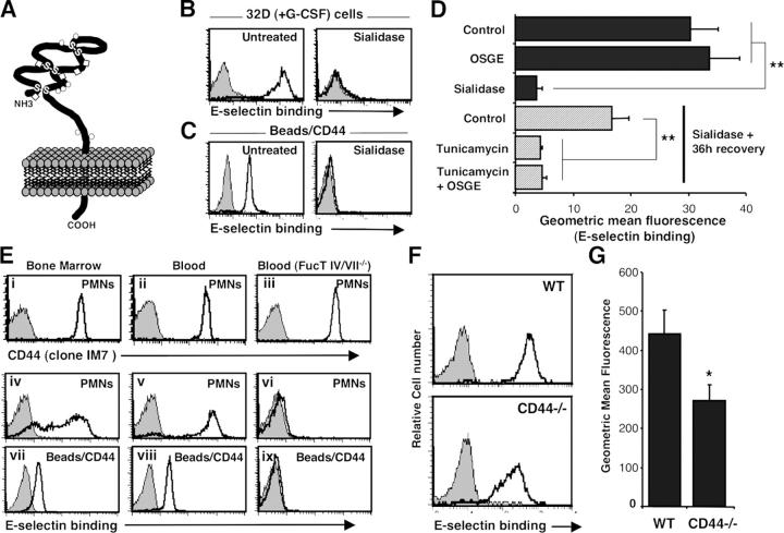
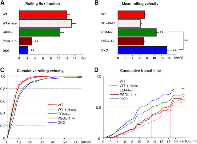
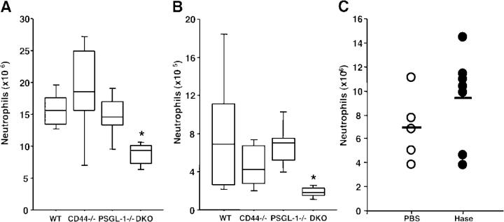
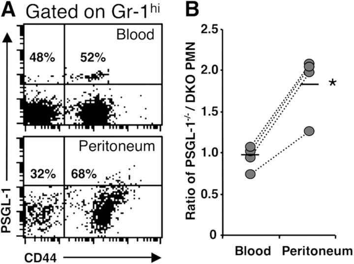
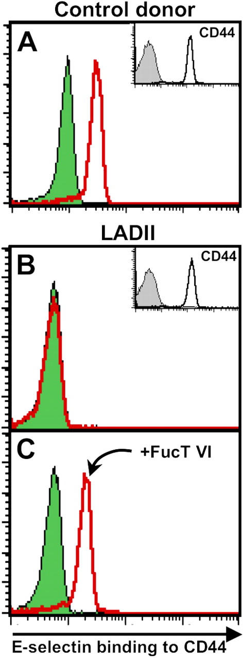
The selectin family of adhesion molecules and their glycoconjugated ligands are essential for blood polymorphonuclear neutrophil (PMN) extravasation into inflammatory and infectious sites. However, E-selectin ligands on PMNs are not well characterized. We show here that CD44 immunopurified from G-CSF-differentiated 32D cells or from peripheral blood PMNs binds specifically to E-selectin. In contrast, CD44 extracted from bone marrow stromal or brain endothelial cell lines does not interact with E-selectin, suggesting cell-specific posttranslational modifications of CD44. PMN-derived CD44 binding activity is mediated by sialylated, alpha(1,3) fucosylated, N-linked glycans. CD44 enables slow leukocyte rolling on E-selectin expressed on inflamed endothelium in vivo and cooperates with P-selectin glycoprotein ligand-1 to recruit neutrophils into thioglycollate-induced peritonitis and staphylococcal enterotoxin A-injected skin pouch. CD44 extracted from human PMNs also binds to E-selectin. Moreover, we demonstrate that CD44 is hypofucosylated in PMNs from a patient with leukocyte adhesion deficiency type II, suggesting that it contributes to the syndrome. These findings thus suggest broader roles for CD44 in the innate immune response and uncover a potential new target for diseases in which selectins play a prominent role.


Figures

Figure 1.
Figure 1.
Neutrophil CD44 binds to E-selectin through N-linked glycans. (A) Structure of CD44 molecule. The globular amino-terminal domain of CD44 proteins contains several potential O- (circle) or N- (diamond) glycosylation sites (for review see reference 31). (B) Binding of E-selectin–IgM chimera to 32D cells differentiated for 4 d in media containing 10 ng/ml G-CSF. All 32D cells bind to E-selectin (left, open histogram) and binding is abrogated by treatment with EDTA (5 mM; gray-filled histograms) or sialidase treatment (right, open histogram). (C) Binding of E-selectin to CD44 immunopurified from the same 32D cells. (D) Contributions of O- and N-glycans in CD44 binding to E-selectin. Differentiated 32D cells were either treated with OSGE, sialidase, or vehicle before immunopurification of CD44. In the last three bars, ESLs were removed by sialidase and cells were allowed to recover for 36 h in the presence or absence of tunicamycin (15 μg/ml) to inhibit N-glycosylation. Data are average geometric mean values from at least three independent experiments. **, P ≤ 0.003. (E) PMN-derived CD44 is an ESL. PMNs were isolated from (i, iv, and vii) BM and (ii, v, and viii) blood of control mice, or (iii, vi, and ix) blood from FucT IV/VII−/− mice. Upper panels (i–iii) show CD44 expression on PMNs (gated on Gr-1hi). Middle panels (iv–vi) depict E-selectin binding on PMNs (Gr-1hi). Lower panels (vii–ix) show binding of E-selectin on immobilized CD44 extracted from purified PMNs (purity >95%). Gray-filled histograms represent isotype-matched control (CD44 staining) or EDTA treatment (E-selectin binding). (F and G) ESLs on wild-type and CD44−/− PMNs. Blood leukocytes were stained with E-selectin–IgM to determine ESL densities. (F) Histograms of E-selectin binding on Gr-1hi PMNs. (G) Geometric mean fluorescence of E-selectin binding on blood Gr-1hi PMNs. n = 12 mice per group. *, P = 0.02.
Figure 2.
Figure 2.
Intravital microscopy of TNF-α–stimulated cremaster muscle venules. Leukocyte behavior in cremasteric venules was recorded between 150 and 210 min after TNF-α administration for off-line analyses. (A) Rolling flux fraction in wild-type (WT, n = 63 venules), WT treated with hyaluronidase (WT + Hase, n = 51), CD44−/− (n = 79), PSGL-1−/− (n = 50), and DKO (n= 59) mice. (B–D) The velocity of 2,610 rolling leukocytes from the five groups of mice was measured over 2 s (also see Fig. S4). (B) Mean rolling velocities. (C) Cumulative histograms of rolling velocities. (D) Cumulative histograms of transit times calculated per 100 μm of venule segment. Five-group comparisons were analyzed by one-way ANOVA with Bonferroni/Dunn post hoc test. **, P < 0.001 (1% risk level) compared with WT group or between indicated two groups.
Figure 3.
Figure 3.
Neutrophil extravasation into inflammatory sites. (A) Thioglycollate-induced peritonitis. Extravasated PMNs were determined 8h after the i.p. injection of thioglycollate (n = 7). (B) SEA-induced inflammation model. Extravasated PMNs were quantified 6 h after instillation of SEA into preformed skin pouches (n = 6). Data are represented by box-and-whisker plots wherein each box represents an interquartile range (central 50%), the median is shown by the horizontal lines, and vertical lines show the full range of data points. Four-group comparisons were analyzed by one-way ANOVA with Bonferroni/Dunn post hoc test. *, P < 0.0083 (5% risk level) compared with WT group. (C) Hyaluronidase treatment does not affect PMN recruitment. PSGL-1−/− mice were injected i.v. with 20 U of hyaluronidase or PBS before i.p. injection of thioglycollate. Each circle represents the value of an individual PSGL-1−/− mouse. P = 0.24.
Figure 4.
Figure 4.
Competitive recruitment of neutrophils into inflammatory sites. Wild-type recipient mice were lethally irradiated and transplanted with mixture of BM cells from PSGL-1−/−CD44+/+ and PSGL-1−/−CD44−/− mice. 6 wk after transplantation, PMNs were elicited by thioglycollate for 8 h. Blood and peritoneal exudates were analyzed by FACS. (A) Representative dot plots for CD44 and PSGL-1 in Gr-1hi cells from blood and peritoneum. (B) Ratios PSGL-1−/−CD44+/+ over PSGL-1−/−CD44−/− Gr-1hi cells. Bars show mean values. n = 4, *, P < 0.01.
Figure 5.
Figure 5.
Human neutrophil CD44 binds to E-selectin. PMNs were purified from healthy donors or from a patient with LADII. PMNs were treated with sialidase or incubated with vehicle. (A) CD44 derived from healthy PMNs binds to E-selectin (empty red histogram), and binding is abrogated by sialidase (green-filled histogram). (B) In contrast, there is no difference in E-selectin binding to immunopurified CD44 between sialidase-treated and sham-treated LADII PMNs. (C) Incubation of LADII PMNs with recombinant FucTVI (20 mU/ml, 40 min at 37°C) and GDP-fucose (1 mM) restores the ability of human CD44 to bind to E-selectin.

References

    1. Lowe, J.B. 2003. Glycan-dependent leukocyte adhesion and recruitment in inflammation. Curr. Opin. Cell Biol. 15:531–538. - PubMed
    1. Ley, K. 2003. The role of selectins in inflammation and disease. Trends Mol. Med. 9:263–268. - PubMed
    1. Steegmaier, M., A. Levinovitz, S. Isenmann, E. Borges, M. Lenter, H.P. Kocher, B. Kleuser, and D. Vestweber. 1995. The E-selectin-ligand ESL-1 is a variant of a receptor for fibroblast growth factor. Nature. 373:615–620. - PubMed
    1. Jones, W.M., G.M. Watts, M.K. Robinson, D. Vestweber, and M.A. Jutila. 1997. Comparison of E-selectin-binding glycoprotein ligands on human lymphocytes, neutrophils, and bovine gamma delta T cells. J. Immunol. 159:3574–3583. - PubMed
    1. Montoya, M.C., K. Holtmann, K.R. Snapp, E. Borges, F. Sanchez-Madrid, F.W. Luscinskas, G. Kansas, D. Vestweber, and M.O. de Landazuri. 1999. Memory B lymphocytes from secondary lymphoid organs interact with E-selectin through a novel glycoprotein ligand. J. Clin. Invest. 103:1317–1327. - PMC - PubMed

Publication types