. 2005 Apr 18;201(8):1257-68.
doi: 10.1084/jem.20042028. Epub 2005 Apr 11.

Boosting antitumor responses of T lymphocytes infiltrating human prostate cancers

Affiliations

Boosting antitumor responses of T lymphocytes infiltrating human prostate cancers

Vincenzo Bronte et al. J Exp Med. .

Abstract

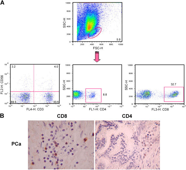
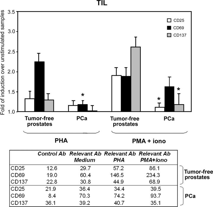
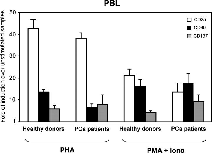
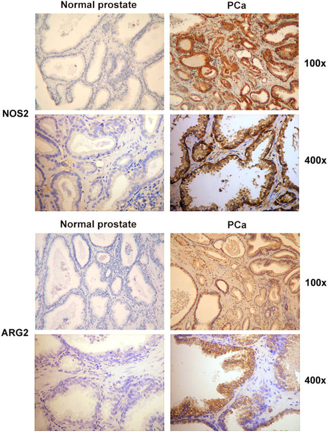
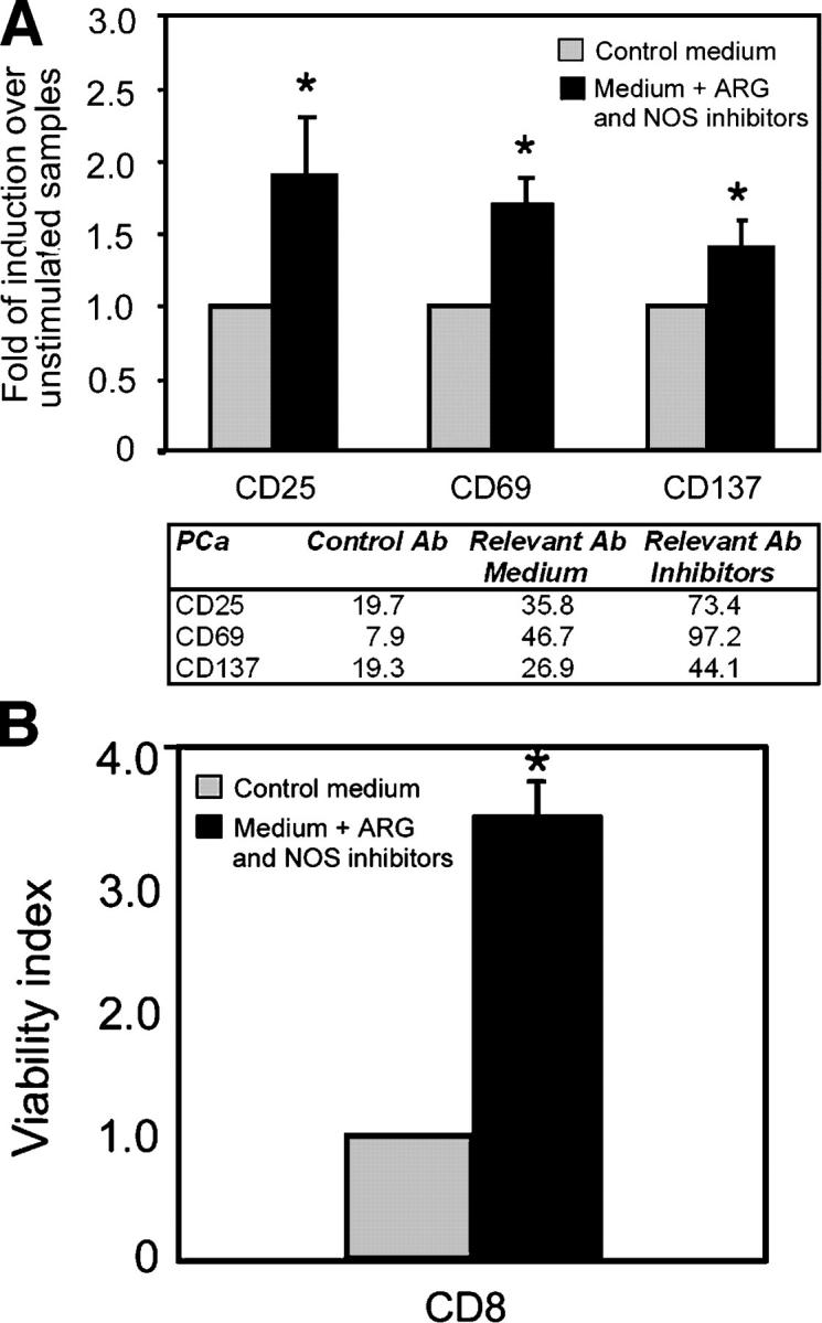
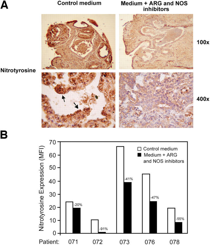
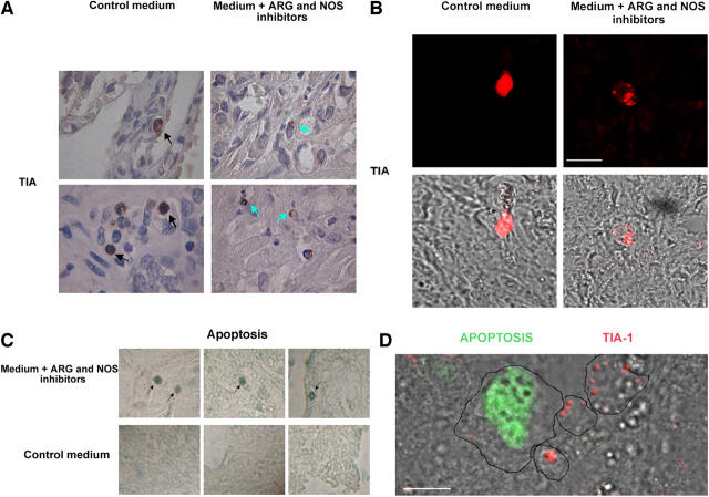
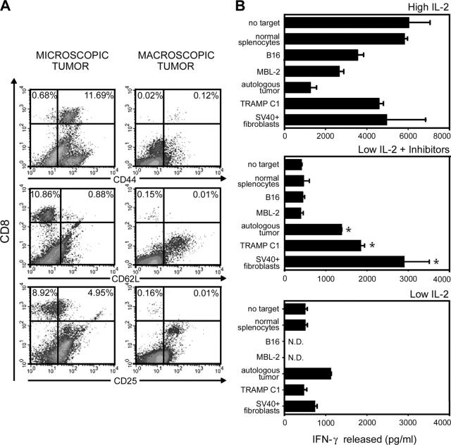
Immunotherapy may provide valid alternative therapy for patients with hormone-refractory metastatic prostate cancer. However, if the tumor environment exerts a suppressive action on antigen-specific tumor-infiltrating lymphocytes (TIL), immunotherapy will achieve little, if any, success. In this study, we analyzed the modulation of TIL responses by the tumor environment using collagen gel matrix-supported organ cultures of human prostate carcinomas. Our results indicate that human prostatic adenocarcinomas are infiltrated by terminally differentiated cytotoxic T lymphocytes that are, however, in an unresponsive status. We demonstrate the presence of high levels of nitrotyrosines in prostatic TIL, suggesting a local production of peroxynitrites. By inhibiting the activity of arginase and nitric oxide synthase, key enzymes of L-arginine metabolism that are highly expressed in malignant but not in normal prostates, reduced tyrosine nitration and restoration of TIL responsiveness to tumor were achieved. The metabolic control exerted by the tumor on TIL function was confirmed in a transgenic mouse prostate model, which exhibits similarities with human prostate cancer. These results identify a novel and dominant mechanism by which cancers induce immunosuppression in situ and suggest novel strategies for tumor immunotherapy.


Figures

Figure 1.
Figure 1.
CD8+ TIL infiltrate malignant prostate. (A) Single-cell suspensions obtained from 50 prostate samples were incubated with antibodies to various T cell markers and analyzed by flow cytometry. A representative experiment is reported. (B) Immunocytochemical detection of CD8+ or CD4+ TIL in PCa tissues (magnification, ×400).
Figure 2.
Figure 2.
TIL in PCa organ cultures are unresponsive. PCa (n = 20) and tumor-free prostate (n = 10) samples were cultured for 48 h in the presence or in the absence of PHA (1 μg/ml) or PMA (50 ng/ml) plus ionomycin (0.5 μg/ml). After enzymatic digestion, the single-cell suspensions obtained from prostate were incubated with antibodies to CD8 plus antibodies to CD25, CD69, and CD137. Flow cytometric analyses were gated on CD8+ lymphocytes. Similar results were obtained when prostate samples were cultured in the presence of IL-2 (100 U/ml). Results are expressed as fold of induction above the mean fluorescence intensity of unstimulated samples, which was taken as 1. *, P < 0.05 compared with tumor-free prostate samples. Mean fluorescence intensity values from a representative experiment are shown in the accompanying table.
Figure 3.
Figure 3.
PBL of PCa patients have normal responses to stimuli. PBL from healthy donors (n = 10) or from the same PCa patients whose prostate samples were analyzed as described in Fig. 2 (n = 20) were cultured for 48 h in the presence or in the absence of PHA (1 μg/ml) or PMA (50 ng/ml) plus ionomycin (0.5 μg/ml). Cells were incubated with antibodies to CD8 plus antibodies to CD25, CD69, and CD137. Flow cytometric analyses were gated on CD8+ lymphocytes. Similar results were obtained when all CD3+ T cells were analyzed. Results are expressed as fold of induction above the mean fluorescence intensity of unstimulated samples, which was taken as 1.
Figure 4.
Figure 4.
Higher expression of NOS2 and ARG2 in PCa tissues than in tumor-free prostatic tissues. Immunohistochemical detection of NOS2 and ARG2 in control (n = 2) and malignant prostatic tissues (n = 4).
Figure 5.
Figure 5.
ARG2 and NOS2 mediate PCa immunosuppression. PCa tissues were cultured 4 d in the presence or in the absence of NOS (l-NMMA; 0.5 mM) plus ARG (NOHA, 0.5 mM) inhibitors. (A) After 4 d of culture, the PCa tissues (n = 33) were digested, and the cell suspensions obtained were incubated with antibodies to CD8 plus antibodies to CD25, CD69, and CD137. Flow cytometric analyses were gated on CD8+ lymphocytes. Results are expressed as fold of induction above the mean fluorescence intensity of untreated samples of the same patient, which was taken as 1 . *, P < 0.05 compared with untreated samples. Mean fluorescence intensity values from a representative experiment are shown in the accompanying table. (B) After 7 d of culture, the PCa tissues (n = 21) were digested, and the cell suspensions obtained were incubated with antibodies to CD8 plus propidium iodide (PI). Data are presented as viability index over untreated tumor cultures of the same patients (number of CD8+ PI cells in treated samples/number of CD8+ PI cells in untreated samples).
Figure 6.
Figure 6.
ARG and NOS inhibitors reduce the level of nitrotyrosines in PCa tissues and in TIL. PCa samples (n = 6) were cultured 4 d in the presence or in the absence of NOS (l-NMMA; 0.5 mM) plus ARG (NOHA; 0.5 mM) inhibitors. (A) Immunohistochemical detection of nitrotyrosines in treated or untreated samples. Arrows indicate highly positive cells present only in untreated PCa tissues. (B) Flow cytometric analysis for nitrotyrosine expression in CD8+ cells infiltrating treated or untreated PCa samples.
Figure 7.
Figure 7.
ARG and NOS inhibitors restore CTL lytic function. (A) Immunohistochemical detection of TIA-1 in PCa samples (n = 10) cultured 4 d in the presence or in the absence of NOS (l-NMMA; 0.5 mM) plus ARG (NOHA; 0.5 mM) inhibitors. The pictures show that in untreated tumor samples cytotoxic granules are evenly distributed in the cytoplasm of CTL (black arrow). In contrast, in tumor samples cultured with inhibitors, CTL cytotoxic granules appear polarized in a confined region of the cell (green arrow: magnification, ×1,000). In each experimental condition, 500 TIA-1+ cells were analyzed. In untreated samples, we counted 23 TIA-1+ cells showing a polarized phenotype (4.6%), whereas in samples cultured in the presence of ARG and NOS inhibitors we counted 461 polarized cells (92.2%). (B) Similar results were obtained by immunofluorescence analysis of TIA-1+ cells. Bar, 10 μm. (C) Immunohistochemical detection of apoptosis by TUNEL assay in PCa samples (n = 10) cultured 4 d in the presence or in the absence of NOS (l-NMMA; 0.5 mM) plus ARG (NOHA; 0.5 mM) inhibitors. Apoptotic cells are indicated (black arrow). In untreated tissues, apoptotic cells were always less than two/sample, whereas in inhibitor-treated tissues we counted >20 apoptotic cells per sample (magnification, ×600). (D) Immunofluorescence analysis of TIA-1+ cells and apoptosis. PCa samples (n = 10) cultured 4 d in the presence of NOS (l-NMMA; 0.5 mM) plus ARG (NOHA; 0.5 mM) inhibitors stained for TIA-1 (red) and subsequently for DNA fragmentation by TUNEL assay (green). Samples were analyzed by confocal microscopy. Bar, 10 μm. Cell margins were drawn to facilitate the readers.
Figure 8.
Figure 8.
ARG and NOS inhibitors are essential to restore full tumor recognition by TIL. (A) Single-cell suspensions obtained from three or four pooled prostate samples from TRAMP mice with small (left) and large (right) tumors, were incubated with antibodies to various T cell markers and analyzed by flow cytometry. A representative experiment is reported. (B) PCa large nodules from three TRAMP mice were enzymatically digested and, after removal of dead cells, were cultured in complete medium alone (to obtain the autologous tumor), in the presence of 100 IU/ml IL-2 (high IL-2), 10 IU/ml IL-2 (low IL-2), 0.5 mM l-NMMA plus 0.5 mM NOHA (inhibitors), or a combination of inhibitors and low-dose IL-2 (low IL-2 plus inhibitors). TIL recovered after 3 wk were used as effectors against a panel of target cells to evaluate the release of IFN-γ after 24-h coincubation. The percentage of CD8+ T lymphocytes in different preparations was greater than 80%. Data are from triplicate wells ± SE. The experiment was repeated twice and, although the cell recovery differed, results of the IFN-γ release assay were comparable. *, P < 0.05 compared with TIL cultured in low IL-2 alone.

References

    1. Finn, O.J. 2003. Cancer vaccines: between the idea and the reality. Nat. Rev. Immunol. 3:630–641. - PubMed
    1. Van Der Bruggen, P., Y. Zhang, P. Chaux, V. Stroobant, C. Panichelli, E.S. Schultz, J. Chapiro, B.J. Van Den Eynde, F. Brasseur, and T. Boon. 2002. Tumor-specific shared antigenic peptides recognized by human T cells. Immunol. Rev. 188:51–64. - PubMed
    1. Rosenberg, S.A., J.C. Yang, and N.P. Restifo. 2004. Cancer immunotherapy: moving beyond current vaccines. Nat. Med. 10:909–915. - PMC - PubMed
    1. Coulie, P.G., and P. van der Bruggen. 2003. T-cell responses of vaccinated cancer patients. Curr. Opin. Immunol. 15:131–137. - PubMed
    1. Parmiani, G., C. Castelli, P. Dalerba, R. Mortarini, L. Rivoltini, F.M. Marincola, and A. Anichini. 2002. Cancer immunotherapy with peptide-based vaccines: what have we achieved? Where are we going? J. Natl. Cancer Inst. 94:805–818. - PubMed

Publication types

MeSH terms