Optical coherence tomography longitudinal evaluation of retinal nerve fiber layer thickness in glaucoma
- PMID: 15824218
- PMCID: PMC1941777
- DOI: 10.1001/archopht.123.4.464
Optical coherence tomography longitudinal evaluation of retinal nerve fiber layer thickness in glaucoma
Erratum in
- Arch Ophthalmol. 2005 Sep;123(9):1206
Abstract
Objectives: To longitudinally evaluate optical coherence tomography (OCT) peripapillary retinal nerve fiber layer thickness measurements and to compare these measurements across time with clinical status and automated perimetry.
Methods: Retrospective evaluation of 64 eyes (37 patients) of glaucoma suspects or patients with glaucoma participating in a prospective longitudinal study. All participants underwent comprehensive clinical assessment, visual field (VF) testing, and OCT every 6 months. Field progression was defined as a reproducible decline of at least 2 dB in VF mean deviation from baseline. Progression of OCT was defined as reproducible mean retinal nerve fiber layer thinning of at least 20 mum.
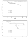
Results: Each patient had a median of 5 usable OCT scans at median follow-up of 4.7 years. The difference in the linear regression slopes of retinal nerve fiber layer thickness between glaucoma suspects and patients with glaucoma was nonsignificant for all variables; however, Kaplan-Meier survival curve analysis demonstrated a higher progression rate by OCT vs VF. Sixty-six percent of eyes were stable throughout follow-up, whereas 22% progressed by OCT alone, 9% by VF mean deviation alone, and 3% by VF and OCT.
Conclusions: A greater likelihood of glaucomatous progression was identified by OCT vs automated perimetry. This might reflect OCT hypersensitivity or true damage identified by OCT before detection by conventional methods.
Figures


References
-
- Sommer A, Pollack I, Maumenee A. Optic disc parameters and onset of glaucomatous field loss. I. Methods and progressive changes in disc morphology. Archives of Ophthalmology. 1979;97:1444–8. - PubMed
-
- Pederson J, Anderson D. The mode of progressive disc cupping in ocular hypertension and glaucoma. Archives of Ophthalmology. 1980;98:490–5. - PubMed
-
- Sommer A, Quigley H, Robin A. Evaluation of nerve fiber layer assessment. Archives of Ophthalmology. 1984;102:1766–71. - PubMed
-
- Sommer A, Katz J, Quigley HA, et al. Clinically detectable nerve fiber atrophy precedes the onset of glaucomatous field loss. Arch Ophthalmol. 1991;109:77–83. - PubMed
-
- Quigley HA, Katz J, Derick RJ, Gilbert D, Sommer A. An evaluation of optic disc and nerve fiber layer examinations in monitoring progression of early glaucoma damage. Ophthalmology. 1992;99:19–28. - PubMed
Publication types
MeSH terms
Grants and funding
LinkOut - more resources
Full Text Sources
Other Literature Sources
Medical
Miscellaneous

