. 2005 Apr 19;102(16):5703-8.
doi: 10.1073/pnas.0500617102. Epub 2005 Apr 11.

Mutations in PTEN-induced putative kinase 1 associated with recessive parkinsonism have differential effects on protein stability

Affiliations

Mutations in PTEN-induced putative kinase 1 associated with recessive parkinsonism have differential effects on protein stability

Alexandra Beilina et al. Proc Natl Acad Sci U S A. .

Abstract

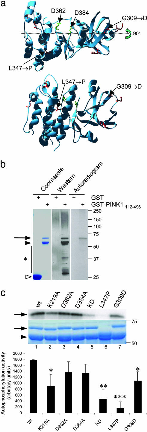
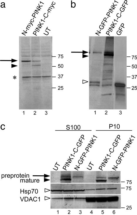
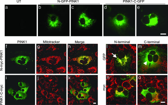
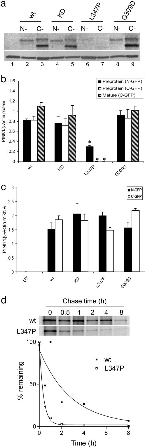
Several mutations in PTEN-induced putative kinase 1 (PINK1) gene have been reported to be associated with recessive parkinsonism. The encoded protein is predicted to be a Ser/Thr protein kinase targeted to mitochondria. In this study, we have investigated the effects of mutations on PINK1 kinase activity in vitro and on expression levels and localization in mammalian cells. We chose to examine two point mutations: G309D, which was originally reported to be stable and properly localized in cells and L347P, which is of interest because it is present at an appreciable carrier frequency in the Philippines. We were able to confirm kinase activity and produce artificial "kinase-dead" mutants that are stable but lack activity. The L347P mutation grossly destabilizes PINK1 and drastically reduces kinase activity, whereas G309D has much more modest effects on these parameters in vitro. This finding is in line with predictions based on homology modeling. We also examined the localization of PINK1 in transfected mammalian cells by using constructs that were tagged with myc or GFP at either end of the protein. These results show that PINK1 is processed at the N terminus in a manner consistent with mitochondrial import, but the mature protein also exists in the cytosol. The physiological relevance of this observation is not yet clear, but it implies that a portion of PINK1 may be exported after processing in the mitochondria.


Figures

Fig. 1.
Fig. 1.
Kinase activity of PINK1 and the effects of mutations in vitro.(a) Model of kinase domain of PINK1 showing positions of critical aspartates and the two recessive mutations investigated in this study. Lower is rotated by 90° at the indicated axis. (b) Expression and autophosphorylation activity of wild-type PINK1 GST-fusion proteins showing, from left to right, Coomassie-stained SDS/PAGE gel, Western blot, and autoradiogram. Alternate lanes contain GST alone, as indicated. Arrow indicates GST-PINK1 fusion proteins, filled arrowhead shows prominent breakdown product, and open arrowhead shows GST alone. Markers on the right of all gels or blots are in kilodaltons. (c) Kinase activity of PINK1 mutants showing autoradiogram (Top), Coomassie staining (Middle), and quantitation of incorporated radioactivity, corrected for protein loading. Wild-type PINK1 (lane 1) is shown for comparison with artificial kinase mutants (K219A, lane 2; D362A, lane 3; D384A, lane 4; triple kinase-dead mutant, lane 5). Two recessive mutants associated with human parkinsonism, L347P and G309D, are shown in lanes 6 and 7. (Bottom) Bars show the average from three independent experiments, with error bars showing SEM. Differences in activity between variants were assessed by ANOVA with Fisher's PLSD post hoc test; *, P < 0.05; **, P < 0.01; ***, P < 0.001.
Fig. 2.
Fig. 2.
Processing of PINK1 protein in mammalian cells. COS-7 cells were transfected with myc-tagged (a) or GFP-tagged (b) constructs. N-terminal (lane 1) and C-terminal (lane 2) tagged versions were used, and either untransfected cells (lane 3 in a) or GFP-transfected cells (lane 3 in b) were included as controls. Arrows show preprotein (before cleavage of the N terminus), and filled arrowhead indicates mature peptide. Open arrowhead in the GFP constructs shows a possible N-terminal fragment (see Results); *, cross-reactivity seen with myc monoclonal in untransfected cells. (c) Subcellular fractionation. COS-7 cells were transfected with C-terminal (lanes 2 and 5) or N-terminal (lanes 3 and 6) GFP-tagged PINK1 or untransfected (lanes 1 and 4) and cell lysates separated into a 10,000 × g pellet (P10, lanes 4–6) and 100,000 × g supernatant (S100, lanes 1–3) fractions. Blot was reprobed with antibodies to Hsp70 and VDAC1 in Middle and Bottom, respectively (open arrowheads). Data are representative of more than two experiments for each construct. Markers on the right of all blots are in kilodaltons.
Fig. 3.
Fig. 3.
Localization of proteins in transfected cells. (ae) COS-7 cells were transfected with GFP-tagged PINK1 constructs, at either the N terminus (b and c) or the C terminus (d and e) and imaged without fixation or staining. Untransfected cells taken at the same exposure settings are shown as a control in a.(fk) Cells were transfected, N-terminal (fh) or C-terminal (ik) myc-tagged PINK1 and counterstained with MitoTracker. After fixation, myc staining was revealed by using anti-myc monoclonal antibody (green in f and i) and compared with MitoTracker (red in g and h). Merged images are shown in h and k.(lo) GFP-vs. myc-tagged proteins, showing merged images only. The arrow in l indicates the area of overlap between N-GFP-PINK1 and MitoTracker compared with a cell transfected with PINK1-C-GFP where the signals remain separated (arrowhead in m). (Scale bars: 10 μm.) Each image is representative of at least three experiments performed in duplicate for each construct.
Fig. 4.
Fig. 4.
Effects of mutations on PINK1 protein stability in mammalian cells. (a) COS-7 cells were transfected with N-terminal (lanes 2,4,6 and 8) or C-terminal (lanes 3, 5, 7, and 9) GFP-tagged PINK1 variants or left untransfected as a control (lane 1). Steady-state protein levels were decreased for L347P (lanes 6 and 7) but not G309D (lanes 8 and 9) recessive mutant PINK1 compared with wild type (lanes 2 and 3) control or the artificial kinase-dead construct (lanes 4 and 5). The arrow indicates the position of the preprotein, the filled arrowhead shows the mature protein, and the open arrowhead shows the position of β-actin used as a loading control. We quantified both protein (b) and mRNA (c) expression for all constructs from n = 3 experiments. For protein data, we measured the intensity of the preprotein band from the N-terminal construct (filled bars) with both the preprotein (open bars) and mature protein (hatched bars) from the C-terminal construct. *, protein levels were significantly different from wild-type construct (P < 0.01 by ANOVA). (d) Pulse–chase analysis of protein stability. COS-7 cells were transiently transfected with N-terminal GFP-tagged wild-type (Upper) or L347P (Lower) PINK1, labeled with 35S-Met/Cys and chased with unlabeled media for 0–8 h as indicated above each lane. Quantification of this experiment is shown, with curves fitted to a single-phase exponential decay function. Similar estimates of stability were obtained in duplicate experiments.

Similar articles

Cited by

References

    1. Kitada, T., Asakawa, S., Hattori, N., Matsumine, H., Yamamura, Y., Minoshima, S., Yokochi, M., Mizuno, Y. & Shimizu, N. (1998) Nature 392, 605–608. - PubMed
    1. Bonifati, V., Rizzu, P., van Baren, M. J., Schaap, O., Breedveld, G. J., Krieger, E., Dekker, M. C., Squitieri, F., Ibanez, P., Joosse, M., et al. (2003) Science 299, 256–259. - PubMed
    1. Hilker, R., Klein, C., Ghaemi, M., Kis, B., Strotmann, T., Ozelius, L. J., Lenz, O., Vieregge, P., Herholz, K., Heiss, W. D. & Pramstaller, P. P. (2001) Ann. Neurol. 49, 367–376. - PubMed
    1. Broussolle, E., Lucking, C. B., Ginovart, N., Pollak, P., Remy, P. & Durr, A. (2000) Neurology 55, 877–879. - PubMed
    1. Scherfler, C., Khan, N. L., Pavese, N., Eunson, L., Graham, E., Lees, A. J., Quinn, N. P., Wood, N. W., Brooks, D. J. & Piccini, P. P. (2004) Brain 127, 1332–1342. - PubMed

Substances