

Nonmammalian gonadotropin-releasing hormone molecules in the brain of promoter transgenic rats
- PMID: 15824321
- PMCID: PMC556124
- DOI: 10.1073/pnas.0501832102
Nonmammalian gonadotropin-releasing hormone molecules in the brain of promoter transgenic rats
Abstract
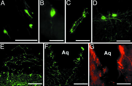
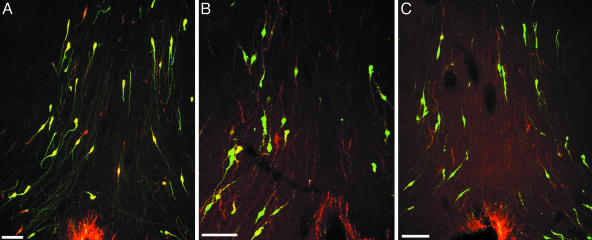
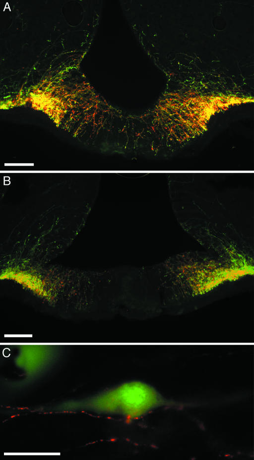
Mammalian gonadotropin-releasing hormone (GnRH1) and nonmammalian immunoreactive GnRH subtypes were examined in transgenic rats carrying an enhanced GFP (EGFP) reporter gene driven by a rat GnRH1 promoter. Double-label immunocytochemistry was performed on EGFP(+)/GnRH1 brain sections by using antisera against GnRH1, GnRH2 (chicken II), GnRH3 (salmon), or seabream GnRH. EGFP(+)/GnRH1 neurons were in the septal-preoptic hypothalamus but not in the midbrain, consistent with GnRH1-immunopositive neurons in WT rats. Apparent coexpression of EGFP(+)/GnRH1 with other GnRH subtypes was observed. All EGFP(+) neurons in the septal-preoptic hypothalamus were GnRH1-immunopositive. However, only approximately 80% of GnRH1-immunopositive neurons were EGFP(+), which awaits further elucidation. GnRH subtypes-immunopositive fibers and EGFP(+)/GnRH1 fibers were conspicuous in the organum vasculosum of the lamina terminalis, median eminence, and surrounding the ependymal walls of the third ventricle and the aqueduct in the midbrain. These results demonstrate that the expression of the EGFP-GnRH1 transgene is restricted to the bona fide GnRH1 population and provide clear morphological evidence supporting the existence of GnRH1 neuronal subpopulations in the septal-preoptic hypothalamus, which might be driven by different segments of the GnRH promoter. This genetic construct permits analyses of promoter usage in GnRH neurons, and our histochemical approaches open questions about functional relations among isoforms of this peptide, which regulates reproductive physiology in its behavioral and endocrine aspects.
Figures
Similar articles
-
Morphological analysis of the early development of telencephalic and diencephalic gonadotropin-releasing hormone neuronal systems in enhanced green fluorescent protein-expressing transgenic medaka lines.J Comp Neurol. 2016 Mar 1;524(4):896-913. doi: 10.1002/cne.23883. Epub 2015 Sep 3. J Comp Neurol. 2016. PMID: 26287569
-
Generation of transgenic rats expressing enhanced green fluorescent protein in gonadotropin-releasing hormone neurons.J Reprod Dev. 2003 Dec;49(6):523-9. doi: 10.1262/jrd.49.523. J Reprod Dev. 2003. PMID: 14967904
-
Localization of immunoreactive lamprey gonadotropin-releasing hormone in the rat brain.Peptides. 1999 Dec;20(12):1503-11. doi: 10.1016/s0196-9781(99)00162-x. Peptides. 1999. PMID: 10698127
-
Electrophysiological characteristics of gonadotrophin-releasing hormone 1-3 neurones: insights from a study of fish brains.J Neuroendocrinol. 2010 Jul;22(7):659-63. doi: 10.1111/j.1365-2826.2010.02035.x. J Neuroendocrinol. 2010. PMID: 20646172 Review.
-
Gonadotropin-releasing hormone neuron development in vertebrates.Gen Comp Endocrinol. 2020 Jun 1;292:113465. doi: 10.1016/j.ygcen.2020.113465. Epub 2020 Mar 14. Gen Comp Endocrinol. 2020. PMID: 32184073 Review.
Cited by
-
Hormone secretion in transgenic rats and electrophysiological activity in their gonadotropin releasing-hormone neurons.Am J Physiol Endocrinol Metab. 2012 Jul 15;303(2):E243-52. doi: 10.1152/ajpendo.00157.2012. Epub 2012 May 22. Am J Physiol Endocrinol Metab. 2012. PMID: 22621869 Free PMC article.
-
Functional significance of GnRH and kisspeptin, and their cognate receptors in teleost reproduction.Front Endocrinol (Lausanne). 2013 Mar 8;4:24. doi: 10.3389/fendo.2013.00024. eCollection 2013. Front Endocrinol (Lausanne). 2013. PMID: 23482509 Free PMC article.
-
Maternal dexamethasone exposure during pregnancy in rats disrupts gonadotropin-releasing hormone neuronal development in the offspring.Cell Tissue Res. 2014 Feb;355(2):409-23. doi: 10.1007/s00441-013-1765-9. Epub 2013 Dec 28. Cell Tissue Res. 2014. PMID: 24374911 Free PMC article.
References
-
- Silverman, A. J., Livne, I. & Witkin, J. W. (1994) in The Physiology of Reproduction, eds. Knobil, E. & Neill, J. D. (Raven, New York), 2nd Ed., pp. 1683–1709.
-
- Jennes, L. & Stumpf, W. E. (1986) Neuroscience 18, 403–416. - PubMed
-
- Merchenthaler, I., Setalo, G., Csontos, C., Petrusz, P., Flerko, B. & Negro-Vilar, A. (1989) Endocrinology 125, 2812–2821. - PubMed
-
- Witkin, J. W. (1990) Neuroscience 37, 501–506. - PubMed
Publication types
MeSH terms
Substances
LinkOut - more resources
Full Text Sources
