

doi: 10.1021/nl050084f.
Six-helix bundles designed from DNA
Affiliations
- PMID: 15826105
- PMCID: PMC3464188
- DOI: 10.1021/nl050084f
Item in Clipboard
Six-helix bundles designed from DNA
Nano Lett.
2005 Apr.
Abstract
We present a designed cyclic DNA motif that consists of six DNA double helices that are connected to each other at two crossover sites. DNA double helices with 10.5 nucleotide pairs per turn facilitate the programming of DNA double crossover molecules to form hexagonally symmetric arrangements when the crossover points are separated by seven or fourteen nucleotide pairs. We demonstrate by atomic force microscopy well-formed arrays of hexagonal six-helix bundle motifs both in 1D and in 2D.
Figures
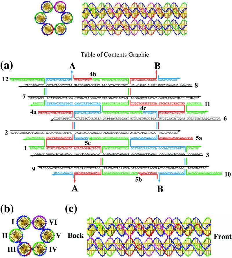
Schematic Drawings of the Six-Helix Bundle Motif. (a) The strand sequences are shown for a version of this motif with 14 nucleotide pairs between crossovers. Points A and B are the places where the cyclic bundle closes. Strand numbering is indicated. There are six helical strands, shown in black, and the other strands perform the crossovers. (b) A cross-sectional geometrical view of a six helix bundle in which the helices are indicated by Roman numerals. (c) A geometrical side view of a six helix bundle in which the crossovers are separated by seven nucleotide pairs. The back and front are indicated as the ends to be used in self-assembly.

Autoradiograms of 5% Non-Denaturing Gels that Establish the Robustness and Proper Stoichiometry of the Six Helix Bundle Motif. Every lane contains the complete blunt-ended 6HB molecule. The lanes are labeled with the radioactive strand that is contained in that complex; strand numbering is as shown in Figure 1. The marker lanes contain a 100-base pair ladder. Note that every strand is incorporated into the motif, and that there are neither multimers nor breakdown products in this gel, demonstrating the robust character of the motif.
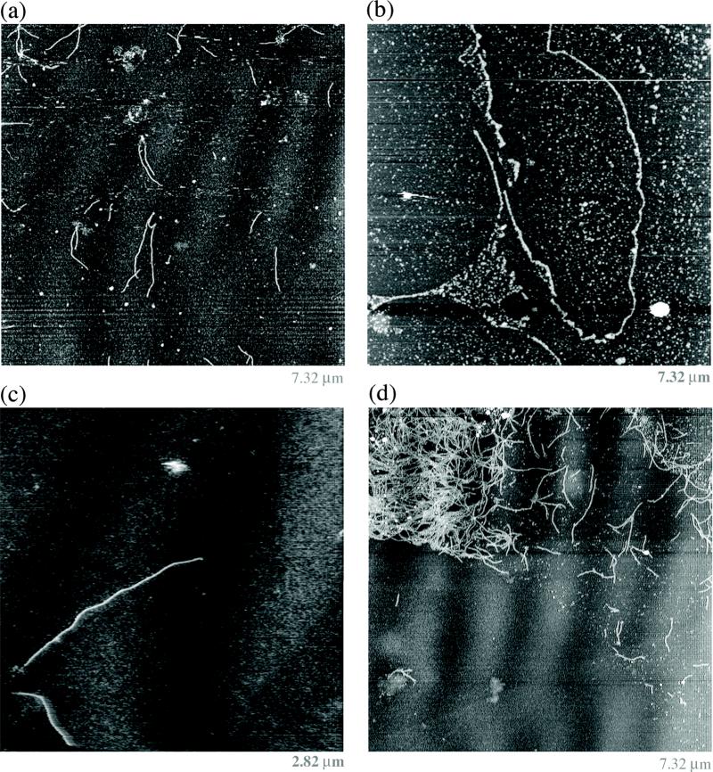
One-Dimensional 6HB Self-Assembled Arrays. The images are obtained by contact mode atomic force microscopy. (a) A series of 6HB wire-like images. (b) A long nanotube. (c) A zoomed view showing a large nanotube. (d) A bundle of nanotubes from which some have been separated. Note that the molecules in all panels are relatively unbent.
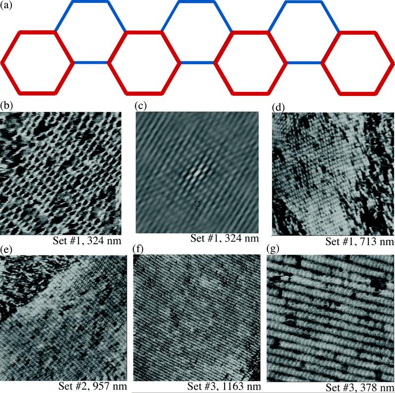
Two dimensional arrays obtained from six helix bundle motifs. The three different paired sets of helical sticky ends have been blunted to produce a 2D array. A schematic where the vertices of hexagons represent helix axes is shown in (a); the red layer is closer to the reader than the blue layer. From the first set, a zoom is shown in panel (b), its autocorrelation function is shown in (c), and a long view is shown in (d). The zoom in (b) shows how the individual 6HB motifs form a corrugated arrangement. Note that the autocorrelation function indicates 2D order. A long view of each of the other two directions is shown in (e) and (f). Neither of their autocorrelation functions indicates good order in more than one direction. (g) shows that the image in (f) has clear vacancies, but that they do not appear to destroy the periodicity of the array.
Similar articles
-
Three-helix bundle DNA tiles self-assemble into 2D lattice or 1D templates for silver nanowires.Nano Lett. 2005 Apr;5(4):693-6. doi: 10.1021/nl050108i. Nano Lett. 2005. PMID: 15826110
-
Prototyping nanorod control: A DNA double helix sheathed within a DNA six-helix bundle.Chem Biol. 2009 Aug 28;16(8):862-7. doi: 10.1016/j.chembiol.2009.07.008. Chem Biol. 2009. PMID: 19716476
-
Atomic force microscopic measurement of the interdomain angle in symmetric Holliday junctions.Biochemistry. 2002 May 14;41(19):5950-5. doi: 10.1021/bi020001z. Biochemistry. 2002. PMID: 11993988
-
Watson-Crick versus Hoogsteen Base Pairs: Chemical Strategy to Encode and Express Genetic Information in Life.Acc Chem Res. 2021 May 4;54(9):2110-2120. doi: 10.1021/acs.accounts.0c00734. Epub 2021 Feb 16. Acc Chem Res. 2021. PMID: 33591181 Review.
-
An overview of structural DNA nanotechnology.Mol Biotechnol. 2007 Nov;37(3):246-57. doi: 10.1007/s12033-007-0059-4. Epub 2007 Jul 12. Mol Biotechnol. 2007. PMID: 17952671 Free PMC article. Review.
Cited by
-
Multi-micron crisscross structures grown from DNA-origami slats.Nat Nanotechnol. 2023 Mar;18(3):281-289. doi: 10.1038/s41565-022-01283-1. Epub 2022 Dec 21. Nat Nanotechnol. 2023. PMID: 36543881 Free PMC article.
-
Metallic nanoparticles used to estimate the structural integrity of DNA motifs.Biophys J. 2008 Oct;95(7):3340-8. doi: 10.1529/biophysj.108.138479. Epub 2008 Jul 11. Biophys J. 2008. PMID: 18621817 Free PMC article.
-
The biological applications of DNA nanomaterials: current challenges and future directions.Signal Transduct Target Ther. 2021 Oct 8;6(1):351. doi: 10.1038/s41392-021-00727-9. Signal Transduct Target Ther. 2021. PMID: 34620843 Free PMC article. Review.
-
Spatially-interactive biomolecular networks organized by nucleic acid nanostructures.Acc Chem Res. 2012 Aug 21;45(8):1215-26. doi: 10.1021/ar200295q. Epub 2012 May 29. Acc Chem Res. 2012. PMID: 22642503 Free PMC article. Review.
-
Synthesis of DNA Origami Scaffolds: Current and Emerging Strategies.Molecules. 2020 Jul 26;25(15):3386. doi: 10.3390/molecules25153386. Molecules. 2020. PMID: 32722650 Free PMC article. Review.
References
Publication types
MeSH terms
Substances
Grants and funding
LinkOut - more resources
Full Text Sources
Other Literature Sources
