. 2005 Aug 1;389(Pt 3):803-12.
doi: 10.1042/BJ20050063.

Secretion of cryptococcal phospholipase B1 (PLB1) is regulated by a glycosylphosphatidylinositol (GPI) anchor

Affiliations

Secretion of cryptococcal phospholipase B1 (PLB1) is regulated by a glycosylphosphatidylinositol (GPI) anchor

Julianne T Djordjevic et al. Biochem J. .

Abstract

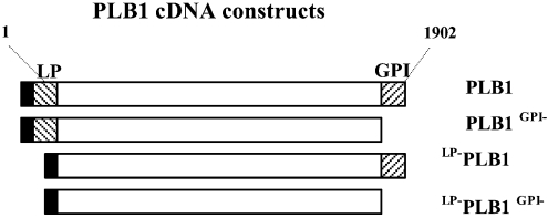
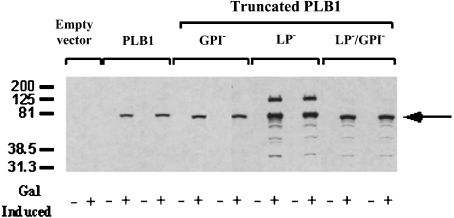
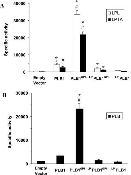
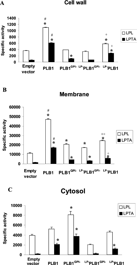
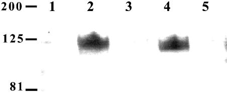
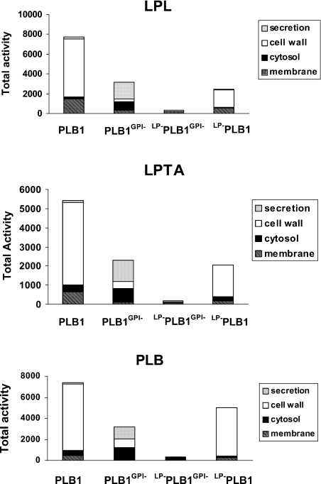
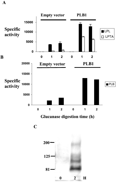
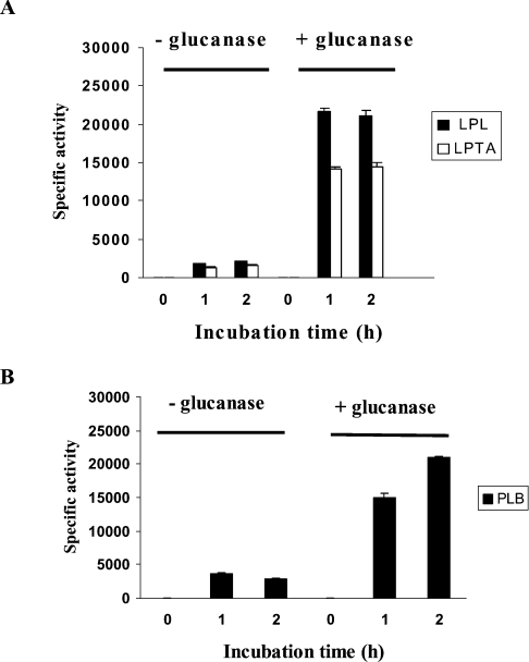
The secreted, multifunctional enzyme PLB1 (phospholipase B1 protein encoded by the PLB1 gene) is a virulence determinant of the pathogenic fungus Cryptococcus neoformans, but the mechanism of its secretion is unknown. The cryptococcal PLB1 gene encodes putative, N-terminal LP (leader peptide) and C-terminal GPI (glycosylphosphatidylinositol) anchor attachment motifs, suggesting that PLB1 is GPI-anchored before secretion. To investigate the role of these motifs in PLB1 secretion, four cDNA constructs were created encoding the full-length construct (PLB1) and three truncated versions without the LP and/or the GPI anchor attachment motifs [(LP-)PLB1 (PLB1 expressed without the LP consensus motif), (LP-)PLB1(GPI-) (PLB1 expressed without the LP and GPI consensus motifs) and PLB1(GPI-) (PLB1 expressed without the GPI anchor attachment motif) respectively]. The constructs were ligated into pYES2, and galactose-induced expression was achieved in Saccharomyces cerevisiae. The LP was essential for secretion of the PLB1 protein and its three activities (PLB, lysophospholipase and lysophospholipase transacylase). Deletion of the GPI motif to create PLB1(GPI-) resulted in a redistribution of activity from the cell wall and membranes to the secreted and cytosolic fractions, with 36-54% of the total activity being secreted as compared with <5% for PLB1. PLB1 produced the maximum cell-associated activity (>2-fold more than that for PLB1(GPI-)), with 75-86% of this in the cell-wall fraction, 6-19% in the membrane fraction and 3-7% in the cytosolic fraction. Cell-wall localization was confirmed by release of activity with beta-glucanase in both S. cerevisiae recombinants and wild-type C. neoformans. The dominant location of PLB1 in the cell wall via GPI anchoring may permit immediate release of the enzyme in response to changing environmental conditions and may represent part of a novel mechanism for regulating the secretion of a fungal virulence determinant.


Figures

Figure 1
Figure 1. Schematic representation of PLB1 cDNA constructs
The N- and C-terminal hatched regions encode the putative 19-amino-acid LP motif (MSIATGTFAFSLFATIAFA) (LP) and the 22-amino-acid GPI anchor attachment motif GAANADVSMGMVALAAGLGLML (GPI) respectively. PLB1 cDNA (1902 bp) was amplified by PCR, incorporating both (PLB1), one (PLB1GPI− or LP−PLB1) or neither (LP−PLB1GPI−) motifs, and each construct was inserted into the pYES2 expression vector. The solid box at the N-terminus represents the His/Express recognition motif that is already present in the pYES2 expression vector.
Figure 2
Figure 2. Detection of intracellular PLB1 expression by Western blotting
PLB1-expressing recombinants (and empty vector control) were cultured in URAgal (+) or URAglu (−) medium to induce and suppress PLB1 expression respectively. Except for the empty vector, two transformants of each recombinant were analysed. Cell lysates were prepared with 0.1% Triton X-100 as described in the Experimental section, and aliquots, containing 30 μg of protein, were subjected to SDS/PAGE and Western blotting. PLB1 constructs were detected with the anti-Express-tagged antibody as described in the Experimental section using ECL and exposure to an X-ray film. The molecular-mass standards (in kDa) are indicated, and the arrow indicates the position of the PLB1 constructs.
Figure 3
Figure 3. Detection of secreted PLB1 by Western blotting
PLB1 recombinants were cultured in URAgal medium to induce PLB1 expression and then allowed to secrete for 22 h. The supernatants (secretions) were collected, and protein (40 μg) was subjected to SDS/PAGE and Western blotting with anti-PLB1 peptide antibody as described in the Experimental section. PLB1 was detected by ECL after exposure to an X-ray film. The molecular mass standards (in kDa) are indicated. Lane 1 contains empty vector control and lanes 2–5 contain PLB1, PLB1GPI−, LP−PLB1 and LP−PLB1GPI− recombinants respectively.
Figure 4
Figure 4. Comparison of secreted PLB1 specific activities by radiometric assay
PLB1 recombinants were cultured in URAgal medium to induce PLB1 expression and then allowed to secrete for 22 h. The supernatants (secretions) were collected and radiometric assays were performed as described in the Experimental section. For LPL/LPTA (A) and PLB (B) assays, 2.5 and 13 μg of protein were used respectively. Results are expressed as mean specific activity±S.E.M. with n=3 [nmol·min−1·(mg of protein)−1 for LPL and LPTA activities and pmol·min−1·mg−1 for PLB activity]. The * indicates that the higher activity relative to the empty vector control is statistically significant (P<0.05). The # indicates that the higher activity for PLB1GPI− relative to PLB1, LP−PLB1 and LP−PLB1GPI− is statistically significant (P<0.05). The + indicates that the higher activity for PLB1 relative to LP−PLB1GPI− and LP−PLB1 is statistically significant (P<0.05). Statistical significance was determined using a paired, two-tailed parametric t test.
Figure 5
Figure 5. Comparison of cell-associated PLB1 specific activities by radiometric assay
PLB1 expression was induced as described for Figure 4. After the 22 h secretion period, the cells were harvested, homogenized and centrifuged at 14000 g. The supernatant was recentrifuged at 100000 g. The 14000 g pellet (cell walls) (A) and the 100000 g pellet (cell membranes) (B) were resuspended in IAB and, together with the 100000 g supernatant (cytosol) (C), were assayed for LPL/LPTA activity. For each fraction, 40, 1 and 2 μg of protein was used respectively. Results are expressed as mean specific activity [nmol·min−1·(mg of protein)−1]±S.E.M with n=3. The * indicates that the higher specific activity relative to the empty vector control is statistically significant (P<0.05). The # indicates that the higher specific activity relative to PLB1GPI−, LP−PLB1 and LP−PLB1GPI− is statistically significant. The + indicates that the higher specific activity relative to PLB1GPI− and LP−PLB1GPI− is statistically significant. The ++ indicates that the higher specific activity relative to LP−PLB1GPI− is statistically significant. For the cytosol, the increased mean PLB1GPI− specific activities compared with the mean PLB1 specific activities are not statistically significant. Statistical significance was determined using a paired, two-tailed parametric t test.
Figure 6
Figure 6. Detection of membrane-associated PLB1 by Western blotting
Total membranes from recombinants were prepared as described for Figure 5 and subjected to SDS/PAGE and Western blotting with anti-PLB1 peptide antibody as described in the Experimental section. PLB1 was detected by ECL after exposure to an X-ray film. The masses of the molecular-mass standards (in kDa) are indicated. Lane 1 contains empty vector control and lanes 2–5 contain PLB1, PLB1GPI−, LP−PLB1 and LP−PLB1GPI− recombinants respectively.
Figure 7
Figure 7. Comparison of the cellular distribution of total PLB1 activity
PLB1 expression was induced as described for Figure 4. After a 22 h secretion period, the cells were pelleted by centrifugation and the supernatants (secretions) were undisturbed. The cell pellets were fractionated into cell wall, membranes and cytosol as described for Figure 5. Each fraction was assayed for LPL, LPTA and PLB activities as described in the Experimental section. The amount of protein used for the LPL/LPTA assays is indicated in Figures 4 and 5. To assay PLB activity, 13, 60, 20 and 20 μg of protein was used for the secreted, cell-wall, membrane and cytosolic fractions respectively. The results were corrected for background from the empty vector control and expressed as the total activity (nmol·min−1·fraction−1 for LPL/LPTA activity and pmol·min−1·fraction−1 for PLB activity).
Figure 8
Figure 8. Analysis of β-glucanase-released proteins from recombinant cell walls
Cell-wall fractions from empty vector control and PLB1-expressing recombinants, prepared as described for Figure 5, were incubated with β-glucanase for 2 h at 37 °C. To assay PLB1 activity released at 0, 1 and 2 h, cell walls were pelleted by centrifugation and aliquots of the supernatant were removed for enzyme assay as described in the Experimental section. For the LPL and LPTA activity assays (A), 1 μl was used in a triplicate assay; for the PLB activity assays (B), 5 μl was used in a duplicate assay. Results are expressed as the mean activity released±S.E.M. with n=3 (nmol·min−1·ml−1 for LPL and LPTA activities and pmol·min−1·ml−1 for PLB activity). Protein was not estimated, due to interference from the added glucanase. The β-glucanase-released proteins were also subjected to SDS/PAGE and Western blotting with an anti-α-agglutinin antibody as described in the Experimental section (C). The GPI-anchored cell-wall marker protein, α-agglutinin, was detected by ECL after exposure to an X-ray film. The masses of the molecular-mass standards (in kDa) are indicated. Lanes 1 and 2 represent 0 and 2 h of β-glucanase digestion respectively.
Figure 9
Figure 9. Release of CnPLB1 activity from cryptococcal cell walls by β-glucanase
Cryptococcal cell walls, prepared as described for Figure 5, were incubated with β-glucanase as described in Figure 8. For the LPL and LPTA activity assays (A), 1 μl was used; for the PLB activity assays (B), 5 μl was used. Results are expressed as the mean activity released±S.E.M. with n=3 (nmol·min−1·ml−1 for LPL and LPTA activities and pmol·min−1·ml−1 for PLB activity). Protein was not estimated due to interference from the added glucanase.

Similar articles

Cited by

References

    1. Chen S. C. A., Wright L. C., Golding J. C., Sorrell T. C. Purification and characterisation of secretory phospholipase B, lysophospholipase-transacylase from a virulent strain of the pathogenic fungus Cryptocococcus neoformans. Biochem. J. 2000;347:431–439. - PMC - PubMed
    1. Cox G. M., McDade H. C., Chen S. C. A., Tucker S. C., Gottfredsson M., Wright L. C., Sorrell T. C., Leidich S. D., Casadevall A., Ghannoum M. A., et al. Extracellular phospholipase activity is a virulence factor for Cryptococcus neoformans. Mol. Microbiol. 2001;39:166–175. - PubMed
    1. Santangelo R., Zoellner H., Sorrell T., Wilson C., Donald C., Djordjevic J. T., Shounan Y., Wright L. C. The role of extracellular phospholipases and mononuclear phagocytes in the dissemination of cryptococcosis in a murine model. Infect. Immun. 2004;72:2229–2239. - PMC - PubMed
    1. Leidich S. D., Ibrahim A. S., Fu Y., Koul A., Jessup C., Vitullo J., Fonzi W., Mirbod F., Nakashima S., Nozawa Y., et al. Cloning and disruption of caPLB1, a phospholipase B gene involved in the pathogenicity of Candida albicans. J. Biol. Chem. 1998;273:26078–26086. - PubMed
    1. Ghannoum M. A. Potential role of phospholipases in virulence and fungal pathogenesis. Clin. Microbiol. Rev. 2000;13:122–143. - PMC - PubMed

Publication types

MeSH terms